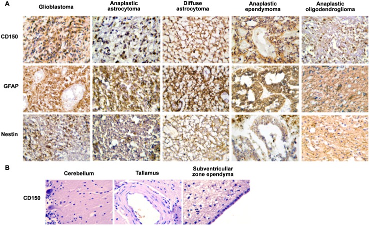
Fig 1. Expression of CD150 in human central nervous system (CNS) tissues.
(A) Immunohistochemical analysis of CD150 expression in human primary CNS tumors. Tumor paraffin sections of glioblastoma, anaplastic astrocytoma, diffuse astrocytoma, anaplastic ependymoma and anaplastic oligodendroglioma were stained with antibodies to CD150 (upper panel), glial fibrillary acidic protein, GFAP, astrocyte marker (middle panel) and nestin, neural stem/progenitor cell marker (lower panel). CD150 expression was detected in all presented histological variants of CNS tumors (upper panel). (B) Expression of CD150 in different regions of human normal brain tissues: cerebellum, thalamus, subventricular zone ependyma. The expression of CD150 was not found in human normal brain tissues. DAB staining shows specific reactions in brown. Cell nuclei are lightly counterstained with haematoxylin. Microscopic magnification of 400× was used for all images.

