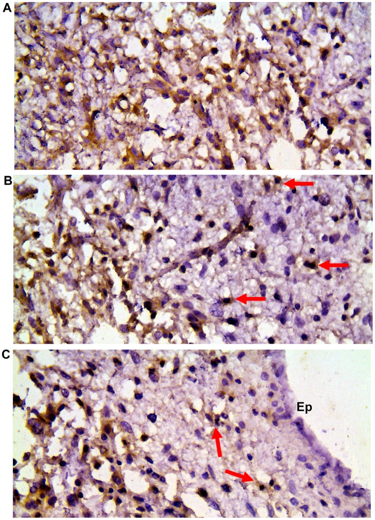
Fig 2. CD150 expression in serial sections of tumor and reactive brain tissues.
(A) Diffuse astrocytoma with extensive microcyst formation. (B) Zone of infiltration with tumor cells of perifocal brain tissue. Scattered tumor cells are shown with arrows. Activated microglia and reactive astrocytes are CD150 negative. (C) Infiltration of subventricular zone with tumor cells (arrows), negative reaction in ependymal cells (Ep). DAB staining shows specific reaction in brown. Cell nuclei are counterstained with haematoxylin. Microscopic magnification 400×.

