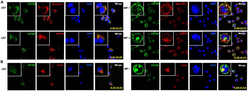
Fig 5. Colocalization of CD150 with the markers of endoplasmic reticulum and Golgi apparatus.
U87 or MP-1 cells were stained for CD150 (green) and the markers of endoplasmic reticulum (A) or Golgi apparatus (B) (red). Markers for endoplasmic reticulum—kinectin-1 and GRP78, for Golgi—furin. Nuclei were visualized by staining with DAPI (4’,6-diamidino-2-phenylindole). Colocalization coefficients were determined using the Manders algorithm (which ranges from 0 to 1, where 0 is defined as no colocalization and 1 as perfect colocalization) and are indicated within the panels. Confocal microscopy shows the similar high colocalisation of CD150 and ER markers in both types of cells, but significantly lower colocalisation of CD150 and Golgi marker in glioma cell line U87 comparing to B cell line MP-1. Microscopic magnification of 630× was used for all pictures. Digital magnification of 3150× was made for the insertions. The data are presented as mean ± SD (n = 7).

