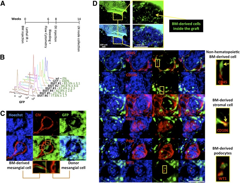
Figure 3.
Host bone marrow-derived cells contribute to mesangial and podocyte regeneration. (A): Overview of experimental plan. Following lethal IR, wt mice were immediately retro-orbitally infused with GFP+ bone marrow cells. Donor engraftment was monitored 6 weeks after transplantation by flow cytometric analysis of the peripheral blood. Following 2 additional weeks, bone marrow chimeric mouse LNs were injected with wt embryonic kidney fragments. Mice were sacrificed 6 weeks later for analysis. (B): Fluorescence intensity profiles of GFP+ leukocytes in peripheral blood of a representative group of bone marrow chimeric mice 6 weeks after transplantation. Blood of a wt mouse and that of a GFP+ mouse were used as negative and positive controls, respectively. (C): Representative 6-week ectopic glomerulus grown inside LNs of GFP bone marrow chimeric mice, showing bone marrow-derived contribution to glomerular mesangium. Sections were stained for CIV (red), and nuclei were counterstained using Hoechst (blue). (D): Pictures of an LN section from a bone marrow chimeric mouse showing localization of GFP+ bone marrow-derived cells in a 6-week kidney graft (upper panels). Representative ectopic glomeruli as in (C) (lower panels). Sections were stained with CD45, CD106, CIV, CD31, Pdpl, or WT1 (red); nuclei were counterstained using Hoechst (blue). Insets show the presence of GFP+CD45−, GFP+CD106+, or GFP+WT1+ cell subsets inside ectopic glomeruli. Abbreviations: BM, bone marrow; BMT, bone marrow transplanted mouse; CIV, collagen IV; EK, embryonic kidney; GFP, green fluorescent protein; IR, irradiation; LN, lymph node; Pdpl, podoplanin; wt, wild-type; WT1, Wilms’ tumor.

