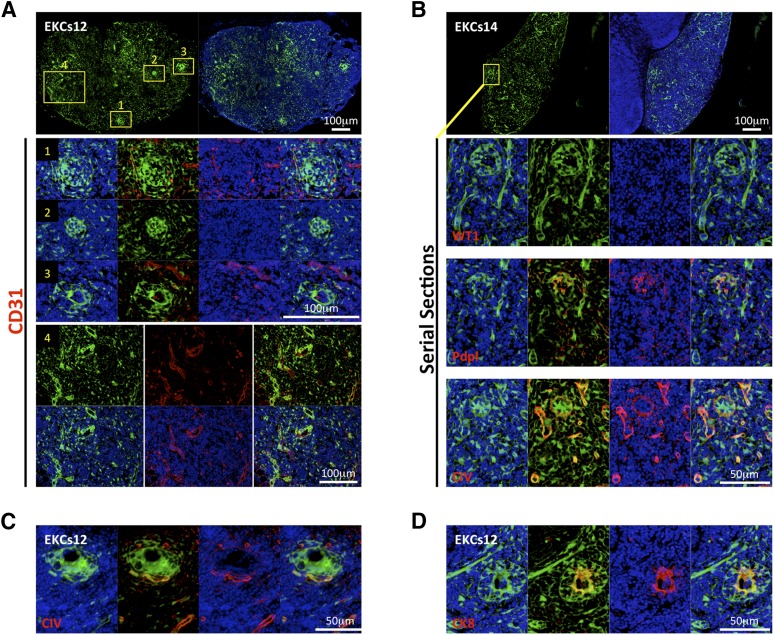
Figure 5.
Whole embryonic kidney single-cell suspensions self-organize into glomerular/nephron-like structures within the LN. (A, B): Pictures of LN sections with the presence of GFP+ (green) donor cells 12 weeks after embryonic kidney single-cell suspension injection; nuclei were counterstained using Hoechst (blue) (upper panels). Enlarged views of the boxed regions of staining for CD31 ([A], middle and lower panels) or WT1, Pdpl, and CIV ([B], lower panels) (red) are shown; nuclei were counterstained using Hoechst (blue). (C, D): Immunofluorescence staining for CIV or CK8 (red) of LN sections as in (A, B). Nuclei were counterstained using Hoechst (blue). Abbreviations: CIV, collagen IV; CK8, cytokeratin-8; EKCs, embryonic kidney cell suspension; Pdpl, podoplanin; WT1, Wilms’ tumor.

