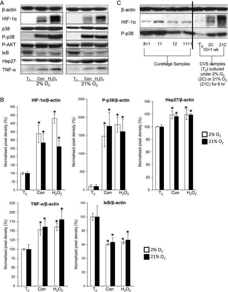
Figure 2.
HIF-1α, p-p38, p38, Hsp27, TNF-α, IκB and P-Akt in first trimester samples (n = 6) cultured under 2% (white bars) or 21% O2 (black bars) in the presence or absence of 1 mM H2O2 for 6 h. (A) Lysates from first trimester explants were immunoblotted with antibodies against HIF-1α, p-p38, p38, Hsp27, TNF-α, IκB and P-AKT and (B) quantified by densitometry. (C) Lysates from representative first trimester curettage samples and a representative CVS sample cultured under 2% O2 (2C) or 21% O2 (21C) were immunoblotted with antibodies against HIF-1α, or phospho-p38. β-Actin staining served to normalize gel loading. Normalized results (±SEM) are plotted, expressing T0 samples as 100%. Significant differences (P < 0.05) are: * versus T0 samples (one-way ANOVA + Student–Newman–Keuls test). Con—denotes samples cultured under a given oxygen concentration for 6 h.

