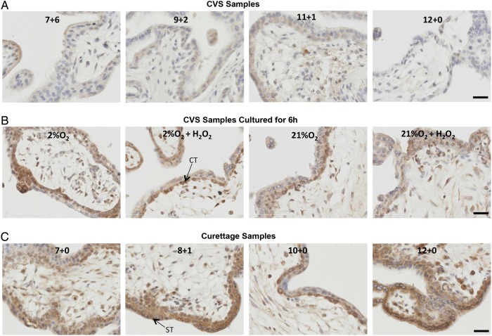
Figure 4.
HIF-2α localization in first trimester placentas collected by a CVS-like technique (A), collected by CVS and then cultured for 6 h (B), or collected by curettage (C). Representative images of HIF-2α staining are shown. Gestational age is indicated in each representative image in A and B, and culture conditions are indicated in C. Brown colour signifies positive staining. HIF-2α was almost undetectable in the CVS tissue (A) whilst a prominent cytotrophoblast and syncytiotrophoblast nuclear staining was detected in all curettage specimens (B) and in cultured explants (C). Scale bar = 50 µm. ST, arrow points to a syncytiotrophoblast nucleus; CT, arrow points to a cytotrophoblast nucleus.

