Figure 4.

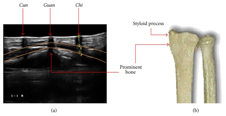
(a) Ultrasonographic image which shows the changes in the vessel features around the pulse measuring locations and (b) it shows the detailed shape of the prominent bone beneath Guan.

(a) Ultrasonographic image which shows the changes in the vessel features around the pulse measuring locations and (b) it shows the detailed shape of the prominent bone beneath Guan.