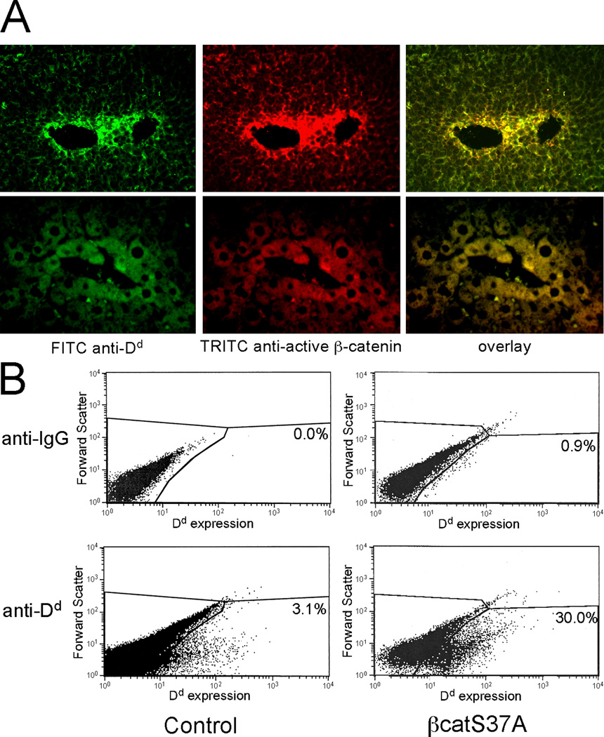
Figure 2. E3-βgl-Dd transgene expression and active β-catenin are co-localized in pericentral hepatocytes.
(A) Transgenic mice containing E3-βgl-Dd were sacrificed at ~8 weeks of age. Sections were stained for Dd expression (left panel, green) and active β-catenin (middle panel, red) as described. Overlay of these (right panel, yellow) demonstrates co-location of Dd and active β-catenin in the same population of hepatoctyes. (B) Hydrodynamic tail-vein injection was used to transfer control plasmid (pcDNA3.1, left panels) and the constitutively active βcatS37A (right panels) into adult E3-βgl-Dd mice. After two days, hepatocytes were isolated, stained with control IgG antibodies (upper two panels) or FITC-anti-Dd antibodies (lower two panels). The percentage of cells gated as positive for Dd expression (those in the lower right area of each panel) are shown.

