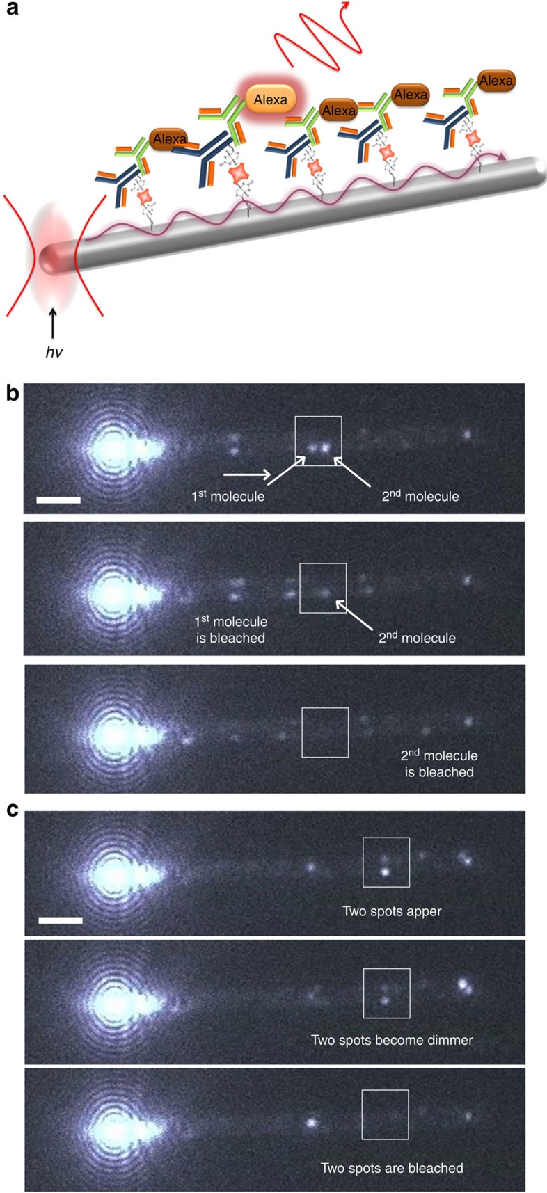
Figure 2. Remote excitation of switching localization microscopy.
(a) Schematic illustration of RE-SFM, the Ag nanowire is irradiated at the left end with focused laser, launching SPPs that propagate along the surface of the nanowire (purple curly arrow), and excite the surface-bound Alexa molecules. (b) RE-SFM images showing two fluorescence spots along the nanowire (enclosed by the white square) bleaching separately. (c) RE-SFM images showing two fluorescent spots along the nanowire (enclosed white square) appearing, dimming and disappearing (blinking off) at the same time in subsequent images. Scale bars, 2 μm.

