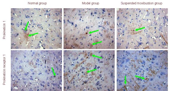
Figure 2.

Prokineticin 1 and prokineticin receptor 1 expression in spinal cord from chronic visceral hypersensitivity rats (immunohistochemistry, × 400). Prokineticin 1 and prokineticin receptor 1 positive cells (stained yellow; arrows) were visible in each group. The expression of positive products was increased in the model group showing dark staining, while staining was light and attenuated in the suspended moxibustion and normal groups.
