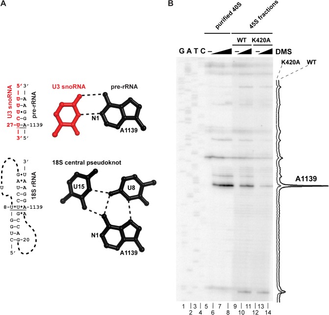
Fig 4. U3 remains base-paired with 18S rRNA in a dhr1 mutant.
(A) Cartoon showing expected U3–18S rRNA base-pairing and the same region of 18S rRNA in the CPK of the mature 40S subunit, based on crystal structure (PDB 3U5B). (B) DMS was used to probe accessibility of A1139. Extracts were prepared from cells expressing WT Dhr1 or Dhr1K420A, as described in the legend to Fig. 2 and fractionated on sucrose density gradients. The 45S regions of the gradients were harvested and treated with DMS as indicated. DMS modification was detected by primer extension using radiolabeled primer AJO1849. Peak areas were quantified using ImageJ (NIH). Purified mature 40S subunits were used as a control and a sequencing ladder was generated using a DNA template containing the 18S rDNA gene.

