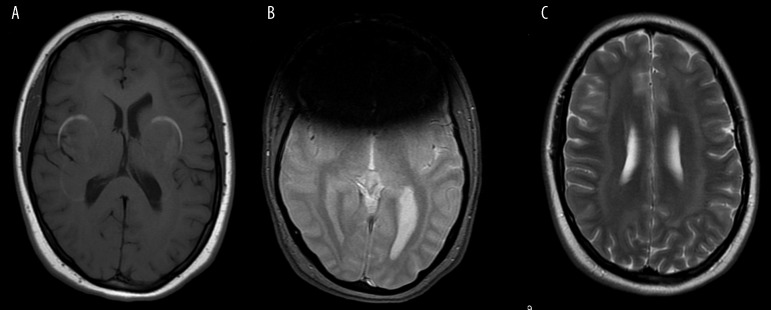
Figure 14A, B.
Orthodontic braces. Typical T1-hyperintense artifacts (A). Loss of signal in GRE/T2*-weighted images (B) makes it impossible to see the anterior part of the brain in this patient with seizures but band heterotopia can be appreciated if the radiologist is familiar with this kind of neuronal migration defect.
Figure 14C. Orthodontic braces. FSE/T2-sequence is less sensitive and band heterotopia can be diagnosed more easily.

