
Fig. 2.
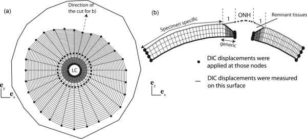
(a) Top view of the surface layer of the finite element (FE) mesh used for the IFEA, showing the nodes at which DIC-measured displacements were applied as kinematic boundary conditions. The outer line represents the location of the most peripheral pixels of the sclera detected by the DIC software. The dashed circle is the border between the generic and specimen-specific mesh. (b) Schematic of the transverse view of the finite element mesh showing the thickness profile in the generic model of the peripapillary sclera. The LC and the remant tissues were not included in the FE model but are represented for clarity. In the inverse analysis, we assumed that the displacements on the surface of the generic peripapillary model were identical to the DIC-measured displacements on the surface of the remnant tissues. The geometry was discretized using mixed Q1P0 trilinear hexahedral elements, with three elements spanning the thickness. The mesh had 40 nodes in the meridional direction and 28 nodes in the circumferential direction. The node density was larger toward the LC to capture the stress concentration due to the compliant tissue of the ONH.