
Figure 5. Pink1 overexpression reduces oxidative stress mediated by thapsigargin.
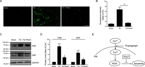
(A and B) ROS probe staining and histograms showing the Pink1 overexpression reduce thapsigargin-induced ROS accumulation in cultured cortical neurons. Results are averages of three independent experiments. Bar=50 μM. Data represent mean±S.E.M. **P<0.01. (C and D) Western blots and histograms showing the HNE and DNP levels are decreased by Pink1 overexpression in cultured cortical neurons. Results are averages of three independent experiments. Data represent mean±S.E.M. *P<0.05 and ***P<0.001. (E) Schematic representation highlighting the protective role of Pink1 in ROS-induced neuronal apoptosis. Ectopic calcium entry by thapsigargin induces oxidative stress and leads to neuronal death. Whereas Pink1 overexpression resists ROS accumulation and protects neurons from apoptosis.