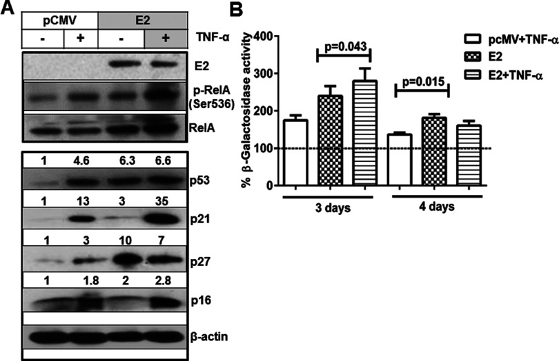
Figure 2. TNF-α-induced NF-κB activation enhanced E2-induced expression of senescent markers.
(A) Whole cell lysates extracted from SiHa cells transfected with pCMV or E2 plasmids (24 h) followed by treatment with TNF-α (20 ng/ml for 24 h) were subjected to immunoblotting studies for the detection of HPV16 E2, p-RelA (Ser536), RelA, p53, p21, p27, p16 and β-actin. Similar results were obtained in another independent experiment. (B) Transfected cells (24 h) as mentioned above were treated with TNF-α for 24 h and after another 24 h (3 days) or 48 h (4 days), lysates were analysed for SA β-galactosidase activity using its substrate (4-MUG) in a fluorimeter with an excitation 360 nm and emission wavelength of 465 nm respectively. The enzyme activity was calculated after normalization against total protein of the lysates and expressed as percentage activity relative to the control-transfected cells. Significance is denoted by P ≤ 0.05.

