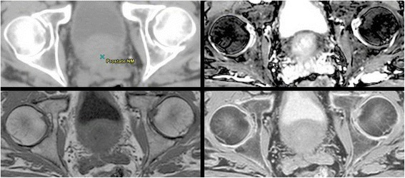
Figure 3.

Different imaging sequences comparing bone boundaries. Detailed Legend: Imaging sequence from top left going clockwise. CT, bTFE, Inverted T1, T1. Notice that boundary of the femoral heads and other bony tissue shows up as a region of no signal in the bTFE and T1 image. On the CT and the inverted T1 image, this edge is readily identifiable.
