Fig. 2.
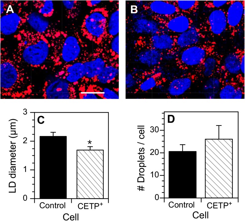
Lipid droplets in vector-transfected SW872 and CETP+ cells. Cells at 70% confluence were incubated with 100 µM oleate/BSA for 24 h to stimulate the development of lipid droplets. Cells were fixed, stained with BODIPY 558/568 C12 (lipid) and DAPI (nucleus), and viewed by confocal microscopy. The number and size of BODIPY 558/568 C12 stained lipid droplets were quantified by ImagePro. A: Vector-transfected SW872 cells. Bar = 10 µm. B: CETP+ cells. C: Quantification of lipid droplet (LD) diameter. D: Number of lipid droplets per cell. Data in C and D (mean ± SD) were derived from analyzing three fields of both cell types, each field containing 83–105 cells. * P < 0.01 versus control cells.

