Fig. 8.
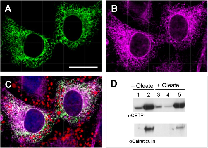
Immunolocalization of CETP. CETP+ cells were grown in the presence of 100 µM oleate/BSA for 48 h to induce lipid droplet formation. A, B: Cells were fixed and immunostained for CETP (A) and calnexin (B). Bar = 20 µm. C: Merge of CETP (green), calnexin (magenta), and BODIPY lipid stain (red). Confocal microscopy images are shown. D: CETP+ cells grown without or with 500 µM oleate were lysed and cellular components fractionated by centrifugation into cytosol (lanes 1 and 4), endoplasmic reticulum-containing membrane (lanes 2 and 5), and lipid droplet (lane 3) fractions. CETP and calreticulin were detected by Western blot.

