Abstract
Respiratory syncytial virus (RSV) infects nearly all children under age 2, and reinfection occurs throughout life, seriously impacting adults with chronic pulmonary diseases. Recent data demonstrate that the anionic pulmonary surfactant lipid phosphatidylglycerol (PG) exerts a potent antiviral effect against RSV in vitro and in vivo. Phosphatidylinositol (PI) is also an anionic pulmonary surfactant phospholipid, and we tested its antiviral activity. PI liposomes completely suppress interleukin-8 production from BEAS2B epithelial cells challenged with RSV. The presence of PI during viral challenge in vitro reduces infection by a factor of >103. PI binds RSV with high affinity, preventing virus attachment to epithelial cells. Intranasal inoculation with PI along with RSV in mice reduces the viral burden 30-fold, eliminates the influx of inflammatory cells, and reduces tissue histopathology. Pharmacological doses of PI persist for >6 h in mouse lung. Pretreatment of mice with PI at 2 h prior to viral infection effectively suppresses inflammation and reduces the viral burden by 85%. These data demonstrate that PI has potent antiviral properties, a long residence time in the extracellular bronchoalveolar compartment, and a significant prophylaxis window. The findings demonstrate PG and PI have complementary roles as intrinsic, innate immune antiviral mediators in the lung.
Keywords: phospholipids, pulmonary surfactant, immunology, inflammation, lung, epithelium, innate immunity, prophylaxis
Respiratory syncytial virus (RSV) is a problematic pathogen for all children before age 2 and is the major cause for hospitalization in newborns (1). Immunity to the virus is incomplete, and there is now clear evidence that RSV infections in adults with chronic lung diseases such as asthma and chronic obstructive pulmonary disease contribute significantly to exacerbation of these underlying conditions (2–6). Infection with RSV can also be fatal in immunocompromised individuals such as bone marrow transplant patients and those with human immunodeficiency virus-acquired immunodeficiency syndrome (7). Worldwide, RSV is estimated to infect 34 million individuals annually and results in high mortality in children during the first 2 years of life (8). The virus is second only to malaria in causing death in early childhood in undeveloped countries (9). Currently, there is no vaccine for the virus, and there are no beneficial medications for treatment of infected individuals.
Pulmonary surfactant is a lipid and protein complex secreted at the air-liquid interface of the alveolar compartment of the lung that plays important roles in reducing alveolar surface tension and regulating innate immunity (10). Recently, several studies have identified significant antiviral activity exerted by the minor anionic pulmonary surfactant phospholipid 1-palmitoyl-2-oleoyl-phosphatidylglycerol (POPG) against respiratory viruses, including multiple strains of RSV and influenza A viruses (11–13). The concentration of POPG and other phosphatidylglycerol (PG) species in the surfactant complex is remarkably high (∼10 mg/ml, 12.5 mM) and greatly exceeds that described for any other tissues. In addition to inhibiting viral infection, POPG also antagonizes the activation of Toll-like receptors (TLRs) 1, 2, 4, and 6 (14, 15). Pulmonary surfactant also contains a second minor anionic phospholipid, phosphatidylinositol (PI), at concentrations of ∼1 mg/ml (∼1.15 mM) (16). Previous work has also identified PI as a regulator of TLRs 1, 2, 4, and 6 (14, 15, 17, 18). The antagonism of TLR4 activation by PI and POPG appears to occur by partially overlapping mechanisms involving the inhibition of CD14 recognition of bacterial lipopolysaccharide (14). These previous observations led us to test whether PI could also inhibit RSV infection in vitro and in vivo. The purpose of this study was to determine whether PI could 1) antagonize RSV-induced inflammatory cytokine production from epithelial cells, 2) inhibit RSV infection in vitro, 3) directly bind to the virus and disrupt cell surface attachment, 4) block RSV infection in mice, and 5) provide prophylaxis against RSV infection. Our findings demonstrate that POPG and PI have overlapping antiviral functions in the lung and suggest that PI or structurally related molecules could be useful in preventing and treating RSV infection.
MATERIALS AND METHODS
Cell culture, viruses, and reagents
HEp2 cells and BEAS2B cells were obtained from American Type Tissue Culture Collection and cultured as previously described (11). The human RSV A2 strain (VR-1540) was also obtained from American Tissue Culture Collection. Virus stocks were propagated in HEp2 cell monolayers using DMEM/Ham’s F12 Medium (GIBCO) plus 5% bovine growth serum (Hyclone) and purified using methods previously described (11, 19, 20). Viral titers and growth were measured using quantitative plaque assays (21). The TLR5 agonist, flagellin, was purchased from Alexis. Agarose was obtained from MP Biomedicals LLC.
Phospholipids
All phospholipids used in these studies were obtained from Avanti Polar Lipids. The biological source of PI was soybean. The molecular species distribution of the PI used in the experiments was determined by MS and consisted of the following molecular ions (mol%): 34:2 (58.4%), 36:2 (14.9%), 36:4 (9.5%), 36:3 (7.8%), 34:3 (6.5%), and 36:5 (1.8%). MS and MS/MS experiments were performed on an AB Sciex QTRAP. An autosampler was used to directly infuse the sample into the mass spectrometer utilizing electrospray ionization in negative ion mode. A methanol-acetonitrile-water (60:20:20, v/v/v) solvent system was used, and soybean PI was diluted to 20 ng/μl. MS/MS experiments utilized a collision energy = 50 eV. The relative intensities of the molecular ion peaks were used to estimate the mol fraction of molecular species within the mass range of 300–900.
In vitro viral infection and phospholipid treatments
BEAS2B cells were seeded at 1.0 × 10⁵/well and grown in 24-well plates for 48 h before the addition of virus. To compare the potency of PI and POPG as antagonists of RSV-elicited inflammatory responses, the cells were challenged with virus at a multiplicity of infection (MOI) of 0.5 in either the absence or presence of 200 μg/ml phospholipids. Phospholipids used in these and all other experiments were bath sonicated until they produced a clear solution, and sterilized by filtration prior to addition to cell cultures. At 48 h after infection, interleukin-8 (IL-8) secretion into the medium was determined by ELISA (11). The phospholipid 1-palmitoyl-2-oleoyl-phosphatidylcholine (POPC) was used as a negative control lipid (11, 12).
To rule out nonspecific effects of lipids on the ability of BEAS2B cells to respond to proinflammatory stimuli, the cells were challenged with the TLR 5 agonist flagellin (10 ng/ml) in either the absence or presence of 200 μg/ml POPG, POPC, or PI. After 48 h, the culture supernatants were harvested, and IL-8 production was measured by ELISA. Additional control experiments were performed to determine the effects of 200 μg/ml phospholipids on BEAS2B protein synthesis, by monitoring the incorporation of 3H leucine into macromolecular pools precipitable by 5% trichloroacetic acid (14)
In additional experiments, we determined the activity of POPG, PI, POPC, and 1,2-dipalmitoyl-phosphatidylcholine (DPPC) as inhibitors of in vitro RSV infection assessed by viral plaque assay. HEp2 cell monolayers (5 × 10⁶ cells/35 mm well) were infected with serial dilutions of virus in the presence or absence of varying concentrations of phospholipid (0–200 μg/ml). After 2 h of viral adsorption, the infecting inoculum and lipids were removed from the wells, which were overlayed with medium containing 0.3% agarose. After 6 days of further incubation, the cultures were fixed and stained, and the viral plaques were quantified (12, 21).
RSV binding to solid-phase phospholipids and HEp2 cells
RSV binding to phospholipids was measured using phospholipid solid phases in microtiter wells, as previously described (11, 13–15). Phospholipids in 100% ethanol were added in 50 μl aliquots (1.5 nmol lipid), to wells in flat-bottomed microtiter plates (Corning). The ethanol was removed under a stream of warm air, and the wells were hydrated and blocked with solutions of 20 mM Tris pH 7.4, 100 mM NaCl, 10 mM CaCl2, containing 3% BSA, and then incubated with varying concentrations of RSV (11, 13). The binding reactions were conducted at 37°C for 2 h. Subsequently, the wells were washed three times with the binding buffer. Specific virus binding was detected with HRP-conjugated anti-human RSV antibody (1:500) (AbD Serotec) by ELISA, using orthophenylenediamine substrate and measuring absorbance at 450 nm (11, 13).
RSV binding to epithelial cell plasma membrane was detected using monolayers of HEp2 cells (2.5 × 10⁵/well in 24-well plates) incubated with varying concentrations of RSV (MOI = 0–10) at 18°C (to minimize endocytosis) for 2 h. The incubations with virus were performed in either the absence or presence of 200 μg/ml phospholipids [POPG, PI, POPC, phosphatidylserine (PS), or phosphatidic acid (PA)]. Following the binding reaction, the monolayers were washed with PBS at 0°C, three times, to remove unattached viruses. The cultures were next harvested by scraping in SDS-PAGE loading buffer and briefly sonicated, and denatured at 95°C, and aliquots were electrophoresed on 8–20% polyacrylamide gels. After electrophoresis, the gels were processed for immunoblotting, and viral proteins were detected with HRP-conjugated anti-human RSV antibody (1:500) using enhanced chemiluminescence (22). The viral G-protein band was chosen for sample comparison, and films were scanned and then analyzed using the National Institutes of Health Image J analysis program (22).
We also conducted phospholipid interaction experiments with RSV to test for the potential viricidal activity of phospholipids. High titer preparations of RSV (1 × 10⁸/ml) were incubated either without or with 200 μg/ml PI or POPC for 1 h at 37°C. Following this incubation, the preparations were diluted 105- to 106-fold and then adsorbed to monolayers of HEp2 cells for 1 h and finally overlayed with 0.3% agarose. After 5 days, the wells were stained, and viral plaques were quantified and compared among the different conditions.
RSV infection in vivo and determination of the PI prophylaxis efficacy window
All experiments involving animals were reviewed and approved by the National Jewish Health Institutional Animal Care and Use Committee. Female BALB/c mice at 6 weeks of age were obtained from Jackson Laboratory. Mice were anesthetized with intraperitoneal Avertin (0.25 g/kg) (11) and either sham infected or infected with RSV [107 plaque forming units (pfu)] in either the absence or presence of 300 μg of PI, or treated with PI alone. The total volume of all intranasal inoculations was kept constant at 50 μl with 25 μl introduced into each nostril.
In a separate series of studies, mice were pretreated with PI to evaluate the activity of this lipid in preventing infection. To determine the efficacy window for PI prophylaxis, mice were anesthetized by isoflurane inhalation and inoculated with PI (600 μg/mouse) at various time points prior to a second round of anesthesia and inoculation with RSV. The prophylaxis studies also included a control group that received PI at the same time as the RSV. For all studies in mice, the experiments were ended at 5 days after infection (the usual peak of infectious virus number) with euthanasia by CO2 asphyxiation. Sample recovery from mice involved lavaging the lungs with 1 ml of saline and dissection and removal of the lungs for tissue processing. The left lungs were harvested and homogenized and processed for RSV plaque assays. The right lungs were used for histopathology scoring (11, 23) by a pathologist who was blinded to the experimental protocol. The lavage material was used to determine total cell infiltrates and quantify different cell populations. The lavage fluid was centrifuged at 200 g × 5 min to remove cells, and the supernatant was used to determine IFN-γ levels (11).
PI turnover in mice
To determine the turnover of instilled PI in vivo, anesthetized mice were inoculated with 150 μg of the lipid in 50 μl of PBS. Subsequently, the mice were euthanized with CO2, and five serial lavages (1 ml each) were performed with buffer (HBSS containing 0.5 mM EGTA and 4.4 mM sodium bicarbonate) at 0, 30, 60, 180, and 360 min after inoculation. Cells were removed by centrifugation at 200 g × 5 min, and the supernatant fluid was processed for lipid extraction as previously described (24). Phospholipid content was determined by measurement of lipid phosphate (25). Phospholipid classes were analyzed by TLC and phosphate determination as previously described (25, 26).
RESULTS
PI inhibits IL-8 production induced by RSV in human epithelial cells
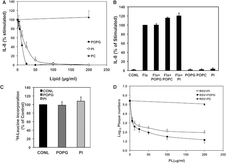
POPG is a minor anionic phospholipid constituent of pulmonary surfactant that has previously been shown to inhibit virus-dependent inflammatory cytokine production (11, 22, 27). In these experiments, we investigated whether PI, another minor anionic phospholipid present in pulmonary surfactant, has similar activity. Initially, we compared PI and POPG as antagonists of RSV-elicited IL-8 production from BEAS2B epithelial cells (11). The cells were pretreated with lipids (200 μg/ml) for 1 h and then infected with RSV (MOI = 0.5), and IL-8 secretion into the culture supernatant was measured at 48 h after infection. As shown in Fig. 1A, both POPG and PI inhibit IL-8 production in a dose-dependent manner. The IC50 value for POPG was 11.4 ± 1.7 (μg/ml), and that of PI was 21.9 ± 5.6 (μg/ml). The control lipid POPC did not have a significant effect on IL-8 production. To exclude any toxic effects of POPG and PI, BEAS2B cells were stimulated with the 10 ng/ml of the TLR5 agonist flagellin, and the response was quantified (11). Neither POPG (200 μg/ml) nor PI (200 μg/ml) significantly inhibited IL-8 production induced by flagellin (Fig. 1B). As an additional control to evaluate lipid toxicity, cultures were incubated with 3H-leucine for 48 h, and the trichloroacetic acid precipitable protein-associated radioactivity was quantified in the absence and presence of the lipid treatment. As shown in Fig. 1C, neither lipid affected protein synthesis measured by 3H-leucine incorporation into macromolecules (11). These data demonstrate that PI disrupts the inflammatory response of BEAS2B cells by a process that does not involve general suppression of TLR signaling, or gross alteration of cellular homeostasis.
Fig. 1.
PI suppresses RSV-induced IL-8 production in BEAS2B cells and inhibits viral propagation in HEp2 cells. IL-8 production by BEAS2B cells that was secreted into the culture supernatant was measured by ELISA (A). Cells were either sham infected or infected with RSV for 48 h at an MOI = 0.5, in either the absence or presence of 200 μg/ml of PI, POPG, or POPC (+PI, +POPG, or +POPC). Values shown are means + SE for three independent experiments. B: IL-8 production in BEAS2B cells was induced with 10 ng/ml of the TLR5 agonist flagellin (Fla) in either the absence or presence of lipids (+POPG, 200 μg/ml; +PI, 200 μg/ml; +POPC, 200 μg/ml), and IL-8 secretion was detected by ELISA. Control (CONL) conditions consisted of sham treatment of the cultures. Additional control conditions show the effects of treatment with lipid alone. C: BEAS2B cells were incubated for 48 h with 3H-leucine, in either the absence or presence of 200 μg/ml POPG, or PI, as indicated. 3H-leucine incorporation into macromolecules precipitated by trichloroacetic acid was measured by liquid scintillation spectrometry. Normalized values shown are means ± SE for three independent experiments. D: HEp2 cells were cultured in 6-well plates and infected with a series of dilutions of RSV (2.5 × 10⁻¹ to 2.5 × 10⁻⁵ dilutions) in either the absence or presence of phospholipids (PL), 0–200 μg/ml of POPC, POPG, or PI, for 2 h. Subsequently, the infecting inocula and lipids were removed, and 0.3% agarose was added in each well. Plaque quantification was performed after 6 days. Plaque numbers shown are means ± SE for three individual experiments.
PI inhibits RSV infection in vitro
In the next series of experiments, we tested the activity of PI as an inhibitor of RSV infection. The antiviral activity of PI was quantified by infecting HEp2 cells for 2 h in either the absence or presence of the indicated concentrations of PI. Subsequently, the viral inoculum and lipids were removed from the cultures, and the cell monolayers were overlayed with agarose. After 5 days, the appearance of plaques was quantified. In Fig. 1D, the data demonstrate that both PI and POPG potently reduce viral plaque formation in a concentration-dependent manner. The activity of PI is comparable to that of POPG, which was previously shown to potently inhibit RSV infection and replication (11). At concentrations of 200 μg/ml, PI reduces viral infection in vitro by a factor >103. These data demonstrate that PI, like POPG, strongly suppresses RSV infection in vitro. We also performed additional experiments examining 200 μg/ml DPPC as a potential inhibitor of viral infection because this phospholipid is the most abundant in pulmonary surfactant. The findings from two independent experiments demonstrated that DPPC did not have any significant inhibitory activity upon RSV plaque formation (virus alone = 3 × 10⁷ pfu/ml, virus + DPPC = 3.3 × 10⁷ pfu/ml, virus + POPG = 3 × 10³ pfu/ml).
PI binds to RSV with high affinity and blocks viral attachment to epithelial cells
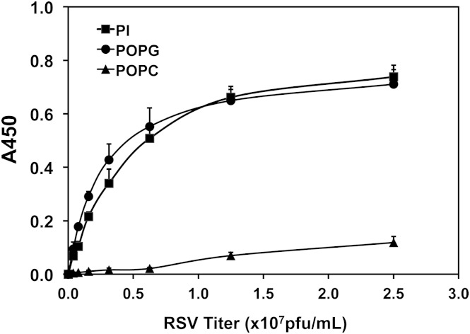
We next sought to elucidate the mechanism by which PI inhibits RSV infection. Previous studies with POPG demonstrated this lipid disrupts RSV infection by direct, high-affinity interactions with the virus that block attachment to epithelial cell plasma membranes (11). We performed solid-phase binding studies to test for direct interactions between RSV and PI, and the results are shown in Fig. 2. RSV bound to solid-phase PI with high affinity and in a saturable reaction, yielding half-maximal binding at a viral titer of 0.25 × 10⁷ pfu/ml. The interaction of the virus with PI was similar to that for POPG. In contrast, the binding of RSV to solid-phase POPC was low affinity and nonspecific.
Fig. 2.
PI binds to RSV with high affinity. Aliquots of 1.5 nmol phospholipid (PI, POPG, or POPC) were adsorbed onto microtiter wells, which were subsequently blocked with 3% albumin in binding buffer. Varying concentrations of RSV (0.05–2.5 × 10⁷ pfu/ml) were added to the wells and incubated at 37°C for 2 h. The wells were washed three times, and viral binding was detected by ELISA using an HRP-conjugated polyclonal antibody and quantified by spectrophotometry at 450 nm. Values shown are means ± SE for three independent experiments.
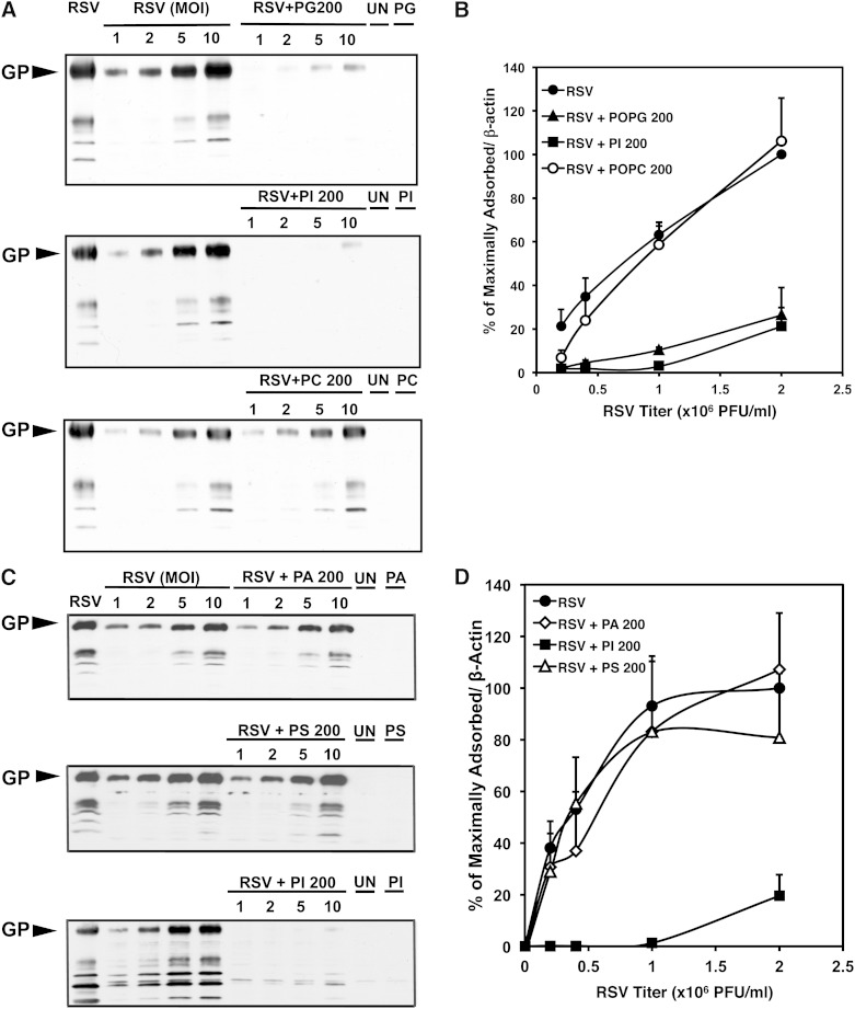
In additional experiments, we examined the activity of PI as an antagonist of RSV attachment to monolayers of HEp2 cells, using Western blotting to detect viral antigens. The experiments were performed at 18°C to block internalization of the virus. For comparison, we also quantified the inhibition of virus binding by other anionic phospholipids including POPG, PA, and PS, and the zwitterionic lipid, POPC. Representative Western blots are shown in Fig. 3A, C, and quantitative data are shown in Fig. 3B, D. The results show that RSV binds to HEp2 cells in a concentration-dependent reaction, and PI effectively disrupts virus attachment to epithelial cells; whereas PA, PS, and POPC do not significantly alter viral attachment. The activity of PI as an antagonist of viral attachment to cells is comparable to that observed for POPG. The data also reveal that the inhibition of RSV binding to cells can be overcome as the viral titer is increased, consistent with PI and POPG exerting a competitive inhibition of the process. From these data, we conclude that specific structural aspects of POPG and PI confer binding affinity and specificity that is not duplicated by either the anionic character, or hydrophobic constituents of other negatively charged lipids, or the hydrophobic constituents of zwitterionic phospholipids (12, 13).
Fig. 3.
PI inhibits RSV binding to HEp2 cells. A: Western blot analysis of RSV binding to monolayers of HEp2 cells. Monolayers of HEp2 cells were incubated with varying concentrations (MOI = 1, 2, 5, and 10) of RSV for 2 h at 18°C in either the absence (RSV) or presence (RSV + PG 200; RSV + PI 200; RSV + PC 200) of 200 μg/ml of lipid. After the binding interval, the monolayers were washed three times with PBS, and then the entire content of each well was harvested and processed by SDS-PAGE and Western blotting. Each blot contains an RSV standard (RSV ∼104 PFU) and lanes corresponding to uninfected cell monolayers (UN) and to monolayers exposed to lipid but no RSV (PG, PI, PC). The location of the viral attachment glycoprotein (GP) is indicated in the left margin. The data in A show a blot from one experiment. B: Summary from three independent experiments performed as described in A and normalized to the amount of β-actin recovered from each well containing HEp2 cells. Values shown are means ± SE. C: Western blot analysis of lipid inhibition of RSV cell surface binding, comparing the activity of PI to that of PA and PS. The experiment was performed as described in A except that the lipids examined were 200 μg/ml PA, PS, or PI. D: Summary of three independent experiments performed as in C and normalized to the amount of β-actin recovered from each well containing HEp2 cells. Values shown are means ± SE.
PI does not act as a viricidal agent
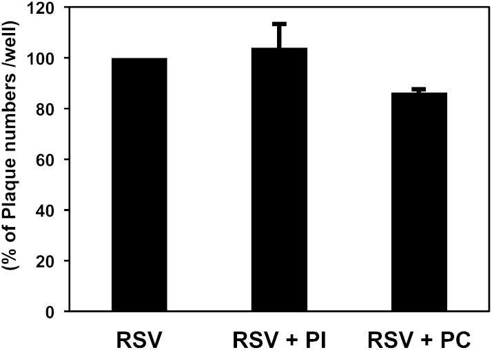
The data in Fig. 3 demonstrate high-affinity interactions between RSV and PI that disrupt viral attachment to epithelial cells but do not eliminate viricidal effects as an additional mechanism of lipid action. We designed experiments to test whether PI could compromise the integrity of RSV and if this could also contribute to the mode of action of the lipid. To examine the inhibitory effect of PI against RSV in more detail, we tested for the reversibility of PI binding to RSV and potential viricidal effects of the phospholipid. High titer preparations of RSV (1 × 10⁸/ml) were incubated with either PI or POPC (200 μg/ml) at 37°C for 1 h. Subsequently, the solutions were diluted 105-fold, and aliquots were analyzed by plaque assay. As shown in Fig. 4, there were no significant differences in the number of recovered plaques with the lipid treatments. The data in Figs. 3 and 4 clearly establish that the principal mode of action of PI as an antagonist of RSV infection is by inhibition of viral attachment to cell surfaces, and not compromise of virus integrity.
Fig. 4.
PI does not inactivate RSV. High titer aliquots of RSV (1 × 10⁸/ml) were incubated either without lipid (RSV) or with 200 μg/ml PI (RSV + PI) or 200 μg/ml PC (RSV + PC) for 1 h at 37°C. Subsequently, the virus was diluted 103- to 106-fold, and the infectivity of the virus was measured by plaque assay as a function of phospholipid (PL) concentration. Values are means ± SE for three independent experiments.
PI suppresses RSV infection in vivo
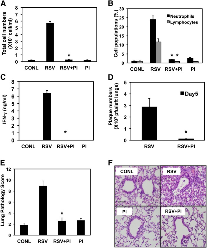
We next conducted experiments to evaluate the effectiveness of PI as an antagonist of RSV infection in vivo. Mice were inoculated with RSV (1 × 10⁷ pfu/mouse) in either the absence or presence of PI (300 μg/mouse), and the progress of viral infection was evaluated after 5 days by quantifying inflammatory cell infiltrates recovered in lavage, IFN-γ production, and the viral burden of RSV within the lung (11, 13). As shown in Fig. 5A, total cell numbers present in bronchoalveolar lavage fluid (BALF) increased ∼25-fold with RSV infection, but simultaneous treatment with RSV plus PI reduced the cell numbers to those of sham-infected animals (CONL = 2.3 ± 0.1 × 10⁴ cells/ml, RSV = 58 ± 2.0 × 10⁴ cells/ml, RSV + PI = 3.0 ± 0.1 × 10⁴ cells/ml, PI alone = 2.5 ± 0.2 × 10⁴ cells/ml). The major virus-elicited, inflammatory cell populations in the BALF were neutrophils and lymphocytes (Fig. 5B), and the presence of both of these cell types was markedly diminished by the PI treatment. High levels of IFN-γ (6.5 ± 0.5 ng/ml) are produced in response to active RSV infections as shown in Fig. 5C, but PI suppression of viral infection and inflammation resulted in no detectable IFN-γ in the BALF (11). We examined the RSV burden present in the left lungs of mice by plaque assay, as previously described (11, 21). The results shown in Fig. 5D demonstrate that PI reduced viral titers by a factor of ∼30. As a control condition, in these infection experiments we also inoculated mice with PI alone, and the results in Fig. 5A–C demonstrate that this treatment was without a significant effect on any of the measured parameters.
Fig. 5.
PI inhibits RSV infection in mice. Balb/C mice (5–8 per group) were anesthetized and inoculated intranasally with 50 μl PBS containing no additions (CONL), or 1 × 10⁷ RSV (RSV), or 1 × 10⁷ RSV + 300 μg PI (RSV + PI), or PI alone (PI). After 5 days, the mice were euthanized and the lungs were lavaged with 1 ml of saline. The recovered lavage solution was centrifuged to produce a cell pellet and supernatant. The mice were dissected and the lungs excised, and the left lobe was processed for quantifying infectious particles by plaque assay. The right lung was processed for histopathology. A: The total cells recovered from lavage. B: The neutrophil and lymphocyte populations recovered in the cell pellet. C: The IFN-γ levels. D: The RSV plaques recovered in homogenates of the left lung. E: The results of quantitative histopathology analysis of tissue from the right lung. F: A representative section from the right lung. Data are from three independent experiments, and the asterisk indicates P < 0.001 (A–D) and P < 0.01 (E).
We also examined the effects of RSV and PI treatment of the mice by quantitative histopathology using light microscopy, which provides a means for comparing tissue damage and inflammation among the different treatment groups. The data in Fig. 5E demonstrate that PI significantly reduces the histological criteria for pulmonary inflammation and infection. Figure 5F shows representative micrographs of right lung sections for the different experimental conditions applied to the mice. The results show that RSV elicits inflammatory responses and damage to the bronchial and alveolar epithelia that are markedly curtailed by PI treatment. Collectively, the data in Fig. 5 demonstrate that in mice, PI significantly suppresses the effects of RSV infection on the lung.
Supplemental PI has a relatively long turnover time in mouse lung
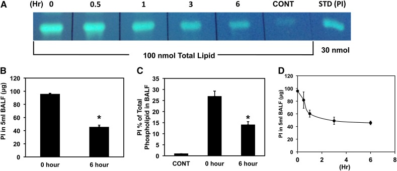
Recently, we reported that supplemental POPG has a relatively short turnover time (t1/2 ∼45 min) in the mouse lung, and this corresponds to a short prophylaxis window for the use of this phospholipid (13). We performed similar turnover studies with PI in mice using a 150 μg (172 nmol) intranasal inoculum. Following administration of PI, groups of mice were euthanized and lavaged after 0, 30, 60, 180, and 360 min, and the amount of PI recovered in the cell-free BALF was measured, using TLC and lipid phosphate determination (13, 25). The data presented in Fig. 6A show that the supplemental PI can be readily recovered and detected in the BALF immediately after the administration. Chemical measurement of PI by phosphate analysis in Fig. 6B demonstrates that the PI turns over relatively slowly with ∼50% of the lipid recoverable at 0 time remaining after 6 h. Figure 6C reveals that the pool of introduced PI recoverable from lavage exceeds that of the endogenous pool by ∼25-fold immediately after instillation and ∼15-fold at 6 h after instillation. Despite these large changes in BALF lipid composition, the levels of PI used in these experiments did not produce any overt signs of distress in the mice, and the histopathology from the experiments described in Fig. 5 also does not show any signs of adverse effects. A plot of the time-dependent loss of PI from the BALF is shown in Fig. 6D and demonstrates an initial rapid turnover followed by slower turnover. The markedly slower turnover phase suggests that PI can persist in the extracellular compartment for extended periods of time.
Fig. 6.
Supplemental PI expands the endogenous surfactant pool and turns over slowly. Balb/C mice (five per group) were given a 50 μl intranasal inoculation of either PBS (CONT) or PBS containing 150 μg of PI. Groups of animals were euthanized at 0, 0.5, 1, 3, and 6 h after the inoculation, and subjected to 5 × 1 ml lavage with saline. Lipids were extracted from the cell-free lavage, separated by TLC, and quantified by phosphate determination. A: Total phospholipids (100 nmol) from each group of mice were separated by TLC, and the lipid classes were visualized by spraying the plates with 8-anilino-napthalene-sulfonate and exposure to UV light. The thin layer plate also contains a 30 nmol PI standard (STD). B: The amount of PI recovered in BALF at 0 h and 6 h. C: The relative amounts of PI expressed as a percentage of the total phospholipids present in cell-free lavage (BALF). D: The turnover of the supplemental PI as a function of time after the intranasal inoculation with the lipid. Data shown are averages from three experiments with five animals per group in each experiment.
PI provides significantly longer prophylaxis against RSV than POPG
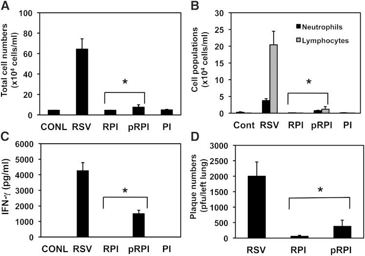
The slower turnover rate of PI compared with that previously reported for POPG (13, 22) prompted us to test whether PI would provide improved protection from RSV infection in mice. In preliminary studies (data not shown), we treated mice with 600 μg of PI at either 6 h or 3 h before RSV infection. The 6 h pretreatment was ineffective, but the 3 h pretreatment reduced viral plaque numbers recovered from the left lungs by ∼50% (data not shown). We next performed in vivo studies using a 600 μg treatment of mice with PI at 2 h prior to infection with RSV, and the results of these experiments are shown in Fig. 7. The RSV infection elicited an expected 14-fold increase in cellular infiltrates recoverable in lavage, which was almost completely suppressed by either simultaneous treatment or pretreatment with PI (Fig. 7A). Likewise, the PI pretreatment reduced the neutrophil and lymphocyte populations to near control levels (Fig. 7B). The data in Fig. 7C demonstrate that the effects of pretreatment with PI upon RSV-elicited IFN-γ levels (75% reduction compared with viral infection alone) were not as pronounced as that observed with simultaneous PI treatment but still demonstrate significant suppression of viral infection. The PI pretreatment reduced the viral burden in the lungs by 85% (Fig. 7D) as compared with the 97% suppression effected by simultaneous treatment with PI. Thus PI prophylaxis provides significant benefit for protecting against RSV infection. Compared with previous prophylaxis studies using POPG, PI more than doubles the efficacy window for protection against the virus.
Fig. 7.
Pretreatment of mice with PI protects against subsequent RSV infection. Balb/C mice (five to eight per group) were anesthetized and inoculated intranasally with 50 μl PBS containing no additions (CONL), or 1 × 10⁷ RSV (RSV), or 1 × 10⁷ RSV + 600 μg PI (RPI), or PI alone (PI). An additional group of mice was anesthetized and pretreated with 600 μg PI 2 h before the RSV treatment (pRPI). After 5 days, the mice were euthanized and the lungs were lavaged with 1ml of saline. The recovered lavage solution was centrifuged to produce a cell pellet and supernatant. The mice were dissected and the lungs excised, and the left lobe was processed for quantifying infectious particles by plaque assay. A: The total cells recovered from lavage. B: The neutrophil and lymphocyte populations recovered in the cell pellet. C: The IFN-γ levels. D: The RSV plaques recovered in homogenates of the left lung. Data are from three independent experiments, and the asterisk indicates P < 0.01 when comparing RSV with either RPI or pRPI conditions.
DISCUSSION
Pulmonary surfactant is a protein- and lipid-rich complex synthesized by alveolar type 2 cells and secreted onto the air/tissue interface of the alveoli (11, 28). Lipids constitute ∼90% of pulmonary surfactant, and proteins comprise ∼10%, by weight. The phospholipid content of surfactant is remarkably high, with concentrations estimated to be 35–50 mg/ml (29, 30). The major lipid class in surfactant is phosphatidylcholine (PC; ∼70% of total phospholipid), and the DPPC molecular species plays a dominant role in reducing alveolar surface tension. The biological role of minor surfactant phospholipid classes, including PG and PI, has been unclear, until recently. Multiple studies now demonstrate the PG and PI act as endogenous suppressors of innate immunity, principally by blocking activation of multiple TLRs (14, 15, 17, 18, 31). This action of PG and PI appears to play a homeostatic role in preventing activation of TLRs by casual exposure to stimuli (e.g., lipopolysaccharide and bacterial membrane constituents) present in the 104 liters of ambient air inspired by adult humans on a daily basis. Thus, the anionic phospholipids set a high threshold for engaging proinflammatory signaling machinery until it is essential. This proposed activity of the anionic surfactant phospholipids complements that also executed by the surfactant proteins A and D (10). The presence of these endogenous inhibitors of pulmonary inflammation, and the tolerance of the lung to high pharmacological doses of these lipids, provides provocative evidence that these lipids can be applied pharmacologically in disease situations where destructive tissue inflammation has spiraled out of control or in the early stages of viral infection.
The studies described in this report further broaden our understanding of the action of pulmonary surfactant PI by demonstrating that this minor phospholipid also can act to neutralize RSV. The in vitro experiments demonstrate that PI prevents RSV from engaging signaling processes required for epithelial cell secretion of IL-8, an early alarm cytokine that serves to recruit neutrophils into the lung. In vitro, the IC50 for PI inhibition of IL-8 secretion is ∼22 μg/ml, a level well below the concentration of the lipid within the alveolar compartment (∼1 mg/ml). One major mechanism by which PI disrupts RSV-elicited proinflammatory signaling is by direct interaction with the virus. Like POPG, PI binds to RSV with high affinity in a concentration-dependent and saturable reaction (11, 22). The high-affinity interaction of PI with RSV blocks the attachment of the virus to epithelial cell plasma membrane and, consequently, infection. Several molecules have been proposed as receptors for RSV including plasma membrane nucleolin and glycosaminoglycans among others (32–35). The inhibition of viral infection in vitro is significant with concentrations of PI of 200 μg/ml reducing infection by a factor of >103.
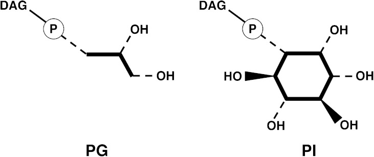
The inhibitory action of PI on RSV attachment to epithelial cells shows structural specificity. Other anionic phospholipids, which are not present in pulmonary surfactant, such as PA and PS, fail to inhibit RSV binding to epithelial cells in vitro. In addition, the zwitterionic phospholipid POPC also fails to alter RSV binding to epithelial cells. Like POPG, the PI molecular species used in this study (derived from soybean) contain unsaturated fatty acids in the diacylglycerol portion of the molecule. However, unsaturated fatty acids are also present in the PS, PA, and PC molecular species used in these experiments, so the dominant determinant appears to reside within the polar head group of the molecule. In molecular modeling (see Fig. 8), the orientation of the phosphodiester bond and two hydroxyls present in the polar head group of PG are structurally superimposable on the phosphodiester bond and hydroxyls in positions 2′ and 3′ of the PI head group ring. The results suggest that anionic character and hydroxyl groups are likely to contribute to the binding interactions between the lipids and virus.
Fig. 8.
Structural comparison between PG and PI head groups. The schematics show common structural features of PG and PI, with dotted lines indicating arrangement of substituents below the plane of the page and dark wedges indicating arrangement of substituents above the plane of the page. DAG denotes the diacylglycerol moiety, and the encircled P indicates a phosphodiester linkage.
The in vitro activity of PI as an antagonist of RSV infection is recapitulated in vivo using a mouse model of viral infection. When PI is introduced at the same time as RSV, using intranasal inoculation, the viral infection is almost completely abrogated. PI dramatically reduces total cellular infiltration and inflammatory cellular infiltration into the lung, reduces the viral burden by a factor of 30, and prevents the engagement of signaling processes required for IFN-γ production. Collectively, these data demonstrate effective neutralization of RSV in vivo and vitro and suggest that if PI can be maintained at sufficient levels within the lung for long enough periods of time, it can be used as a therapeutic for arresting an established viral infection.
We also examined the potential role of PI as a preventative against RSV infection. In Fig. 6, we provide evidence that pharmacological supplementation of mice with PI results in significant expansion of the endogenous pool of the lipid, and this expansion appears relatively long lived. Approximately 50% of the applied PI dose was retained in the extracellular bronchoalveolar compartment for 6 h. This apparently slow turnover of PI corresponded with a significant window of efficacy for the lipid to protect the host from viral infection. When PI was introduced 2 h prior to RSV infection, the total and inflammatory cellular infiltrates in BALF were reduced to near control levels, the IFN-γ levels were reduced by 75%, and the viral burden within the lung was reduced by 85%. Although the 2 h pretreatment with PI afforded significant protection against RSV infection, the infectious particles recovered from the lung and the IFN-γ levels indicate that a modest level of viral infection occurred. This antiviral action of PI is significantly longer than that found for POPG, which had a prophylaxis efficacy window of 45min.
In considering the findings of this study, it is important to also factor in the differences between mouse and human respiratory physiology. Mice breathe at a rate up to 300 times/min, whereas in human newborns the rate is 25 times/min. The turnover of pulmonary surfactant constituents is coupled to the respiratory rate. Perhaps this is best illustrated from stable isotope studies in human neonates supplemented with artificial pulmonary surfactant that reveal that the t1/2 for POPG in the lung is ∼30 h (36). Such data suggest that both POPG and PI administered in pharmacological doses to humans should have significant benefit for both preventing and treating RSV infections.
In summary, our data demonstrate that the minor anionic pulmonary surfactant phospholipid PI has potent antiviral activity against RSV, which is a serious respiratory pathogen, without an effective vaccine for prevention. PI binds the virus with high affinity and prevents its attachment to, and infection of, epithelial cells. Pharmacological doses of PI are relatively long lived in the mouse lung and show better prophylaxis activity than POPG. These findings suggest new pharmacological approaches to both prevention and treatment of RSV in both developed and underdeveloped countries.
Footnotes
Abbreviations:
- BALF
- bronchoalveolar lavage fluid
- DPPC
- 1,2-dipalmitoyl-phosphatidylcholine
- IL-8
- interleukin 8
- MOI
- multiplicity of infection
- PA
- phosphatidic acid
- PC
- phosphatidylcholine
- PG
- phosphatidylglycerol
- PI
- phosphatidylinositol
- POPC
- 1-palmitoyl-2-oleoyl-phosphatidylcholine
- POPG
- 1-palmitoyl-2-oleoyl-phosphatidylglcyerol
- PS
- phosphatidylserine
- RSV
- respiratory syncytial virus
- TLR
- Toll-like receptor
This work was supported by National Institutes of Health Grant HL073907 (D.R.V.) and by the state of Colorado CBDEG-2009 (D.R.V.) and CBDEG-2012 (M.N.).
REFERENCES
- 1.Hall C. B. 2001. Respiratory syncytial virus and parainfluenza virus. N. Engl. J. Med. 344: 1917–1928. [DOI] [PubMed] [Google Scholar]
- 2.Falsey A. R. 2007. Respiratory syncytial virus infection in adults. Semin. Respir. Crit. Care Med. 28: 171–181. [DOI] [PubMed] [Google Scholar]
- 3.Falsey A. R., Formica M. A., Hennessey P. A., Criddle M. M., Sullender W. M., Walsh E. E. 2006. Detection of respiratory syncytial virus in adults with chronic obstructive pulmonary disease. Am. J. Respir. Crit. Care Med. 173: 639–643. [DOI] [PMC free article] [PubMed] [Google Scholar]
- 4.Falsey A. R., Hennessey P. A., Formica M. A., Cox C., Walsh E. E. 2005. Respiratory syncytial virus infection in elderly and high-risk adults. N. Engl. J. Med. 352: 1749–1759. [DOI] [PubMed] [Google Scholar]
- 5.Nicholson K. G., Kent J., Ireland D. C. 1993. Respiratory viruses and exacerbations of asthma in adults. Br. Med. J. 307: 982–986. [DOI] [PMC free article] [PubMed] [Google Scholar]
- 6.Teichtahl H., Buckmaster N., Pertnikovs E. 1997. The incidence of respiratory tract infection in adults requiring hospitalization for asthma. Chest. 112: 591–596. [DOI] [PMC free article] [PubMed] [Google Scholar]
- 7.Whimbey E., Ghosh S. 2000. Respiratory syncytial virus infections in immunocompromised adults. Curr. Clin. Top. Infect. Dis. 20: 232–255. [PubMed] [Google Scholar]
- 8.Nair H., Nokes D. J., Gessner B. D., Dherani M., Madhi S. A., Singleton R. J., O’Brien K. L., Roca A., Wright P. F., Bruce N., et al. 2010. Global burden of acute lower respiratory infections due to respiratory syncytial virus in young children: a systematic review and meta-analysis. Lancet. 375: 1545–1555. [DOI] [PMC free article] [PubMed] [Google Scholar]
- 9.Lozano R., Naghavi M., Foreman K., Lim S., Shibuya K., Aboyans V., Abraham J., Adair T., Aggarwal R., Ahn S. Y., et al. 2012. Global and regional mortality from 235 causes of death for 20 age groups in 1990 and 2010: a systematic analysis for the Global Burden of Disease Study 2010. Lancet. 380: 2095–2128. [DOI] [PMC free article] [PubMed] [Google Scholar]
- 10.Wright J. R. 2005. Immunoregulatory functions of surfactant proteins. Nat. Rev. Immunol. 5: 58–68. [DOI] [PubMed] [Google Scholar]
- 11.Numata M., Chu H. W., Dakhama A., Voelker D. R. 2010. Pulmonary surfactant phosphatidylglycerol inhibits respiratory syncytial virus-induced inflammation and infection. Proc. Natl. Acad. Sci. USA. 107: 320–325. [DOI] [PMC free article] [PubMed] [Google Scholar]
- 12.Numata M., Kandasamy P., Nagashima Y., Posey J., Hartshorn K., Woodland D., Voelker D. R. 2012. Phosphatidylglycerol suppresses influenza A virus infection. Am. J. Respir. Cell Mol. Biol. 46: 479–487. [DOI] [PMC free article] [PubMed] [Google Scholar]
- 13.Numata M., Grinkova Y. V., Mitchell J. R., Chu H. W., Sligar S. G., Voelker D. R. 2013. Nanodiscs as a therapeutic delivery agent: inhibition of respiratory syncytial virus infection in the lung. Int. J. Nanomedicine. 8: 1417–1427. [DOI] [PMC free article] [PubMed] [Google Scholar]
- 14.Kuronuma K., Mitsuzawa H., Takeda K., Nishitani C., Chan E. D., Kuroki Y., Nakamura M., Voelker D. R. 2009. Anionic pulmonary surfactant phospholipids inhibit inflammatory responses from alveolar macrophages and U937 cells by binding the lipopolysaccharide-interacting proteins CD14 and MD-2. J. Biol. Chem. 284: 25488–25500. [DOI] [PMC free article] [PubMed] [Google Scholar]
- 15.Kandasamy P., Zarini S., Chan E. D., Leslie C. C., Murphy R. C., Voelker D. R. 2011. Pulmonary surfactant phosphatidylglycerol inhibits Mycoplasma pneumoniae-stimulated eicosanoid production from human and mouse macrophages. J. Biol. Chem. 286: 7841–7853. [DOI] [PMC free article] [PubMed] [Google Scholar]
- 16.Wright S. M., Hockey P. M., Enhorning G., Strong P., Reid K. B., Holgate S. T., Djukanovic R., Postle A. D. 2000. Altered airway surfactant phospholipid composition and reduced lung function in asthma. J. Appl. Physiol. 89: 1283–1292. [DOI] [PubMed] [Google Scholar]
- 17.Mueller M., Brandenburg K., Dedrick R., Schromm A. B., Seydel U. 2005. Phospholipids inhibit lipopolysaccharide (LPS)-induced cell activation: a role for LPS-binding protein. J. Immunol. 174: 1091–1096. [DOI] [PubMed] [Google Scholar]
- 18.Wang P. Y., Kitchens R. L., Munford R. S. 1998. Phosphatidylinositides bind to plasma membrane CD14 and can prevent monocyte activation by bacterial lipopolysaccharide. J. Biol. Chem. 273: 24309–24313. [DOI] [PubMed] [Google Scholar]
- 19.Dakhama A., Lee Y-M., Gelfand E. W. 2005. Virus-induced airway dysfunction: pathogenesis and biomechanisms. Pediatr. Infect. Dis. J. 24: S159–S169. [DOI] [PubMed] [Google Scholar]
- 20.LeVine A. M., Elliott J., Whitsett J. A., Srikiatkhachorn A., Crouch E., DeSilva N., Korfhagen T. 2004. Surfactant protein-D enhances phagocytosis and pulmonary clearance of respiratory syncytial virus. Am. J. Respir. Cell Mol. Biol. 31: 193–199. [DOI] [PubMed] [Google Scholar]
- 21.McKimm-Breschkin J. L. 2004. A simplified plaque assay for respiratory syncytial virus—direct visualization of plaques without immunostaining. J. Virol. Methods. 120: 113–117. [DOI] [PubMed] [Google Scholar]
- 22.Numata M., Nagashima Y., Moore M. L., Berry K. Z., Chan M., Kandasamy P., Peebles R. S., Jr., Murphy R. C., Voelker D. R. 2013. Phosphatidylglycerol provides short-term prophylaxis against respiratory syncytial virus infection. J. Lipid Res. 54: 2133–2143. [DOI] [PMC free article] [PubMed] [Google Scholar]
- 23.Cimolai N., Taylor G. P., Mah D., Morrison B. J. 1992. Definition and application of a histopathological scoring scheme for an animal model of acute Mycoplasma pneumoniae pulmonary infection. Microbiol. Immunol. 36: 465–478. [DOI] [PubMed] [Google Scholar]
- 24.Bligh E. G., Dyer W. J. 1959. A rapid method of total lipid extraction and purification. Can. J. Biochem. Physiol. 37: 911–917. [DOI] [PubMed] [Google Scholar]
- 25.Rouser G., Siakatos A. N., Fleischer S. 1966. Quantitative analysis of phospholipids by thin layer chromatography and phosphorous analysis of spots. Lipids. 1: 85–86. [DOI] [PubMed] [Google Scholar]
- 26.Chiba H., Pattanajitvilai S., Evans A. J., Harbeck R. J., Voelker D. R. 2002. Human surfactant protein D (SP-D) binds Mycoplasma pneumoniae by high affinity interactions with lipids. J. Biol. Chem. 277: 20379–20385. [DOI] [PubMed] [Google Scholar]
- 27.Numata M., Kandasamy P., Voelker D. R. 2012. Anionic pulmonary surfactant lipid regulation of innate immunity. Expert Rev. Respir. Med. 6: 243–246. [DOI] [PMC free article] [PubMed] [Google Scholar]
- 28.Erpenbeck V. J., Malherbe D. C., Sommer S., Schmiedl A., Steinhilber W., Ghio A. J., Krug N., Wright J. R., Hohlfeld J. M. 2005. Surfactant protein D increases phagocytosis and aggregation of pollen-allergen starch granules. Am. J. Physiol. Lung Cell. Mol. Physiol. 288: L692–L698. [DOI] [PubMed] [Google Scholar]
- 29.Lewis J. F., Jobe A. H. 1993. Surfactant and the adult respiratory distress syndrome. Am. Rev. Respir. Dis. 147: 218–233. [DOI] [PubMed] [Google Scholar]
- 30.Schmidt R., Meier U., Markart P., Grimminger F., Velcovsky H. G., Morr H., Seeger W., Gunther A. 2002. Altered fatty acid composition of lung surfactant phospholipids in interstitial lung disease. Am. J. Physiol. Lung Cell. Mol. Physiol. 283: L1079–L1085. [DOI] [PubMed] [Google Scholar]
- 31.Hashimoto M., Asai Y., Ogawa T. 2003. Treponemal phospholipids inhibit innate immune responses induced by pathogen-associated molecular patterns. J. Biol. Chem. 278: 44205–44213. [DOI] [PubMed] [Google Scholar]
- 32.Hallak L. K., Collins P. L., Knudson W., Peeples M. E. 2000. Iduronic acid-containing glycosaminoglycans on target cells are required for efficient respiratory syncytial virus infection. Virology. 271: 264–275. [DOI] [PubMed] [Google Scholar]
- 33.Tripp R. A., Jones L. P., Haynes L. M., Zheng H., Murphy P. M., Anderson L. J. 2001. CX3C chemokine mimicry by respiratory syncytial virus G glycoprotein. Nat. Immunol. 2: 732–738. [DOI] [PubMed] [Google Scholar]
- 34.Marr N., Turvey S. E. 2012. Role of human TLR4 in respiratory syncytial virus-induced NF-κB activation, viral entry and replication. Innate Immun. 18: 856–865. [DOI] [PubMed] [Google Scholar]
- 35.Tayyari F., Marchant D., Moraes T. J., Duan W., Mastrangelo P., Hegele R. G. 2011. Identification of nucleolin as a cellular receptor for human respiratory syncytial virus. Nat. Med. 17: 1132–1135. [DOI] [PubMed] [Google Scholar]
- 36.Hallman M., Merritt T. A., Bry K. 1994. The fate of exogenous surfactant in neonates with respiratory distress syndrome. Clin. Pharmacokinet. 26: 215–232. [DOI] [PubMed] [Google Scholar]


