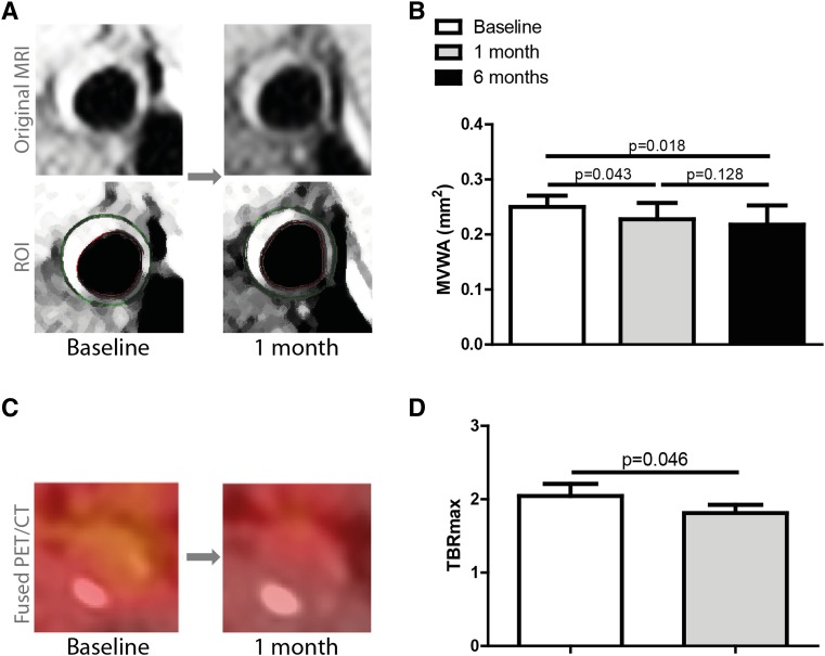
Fig. 4.
Imaging results. MVWA and TBRmax of the carotid arteries, as assessed by MRI and FDG-PET/CT scan, respectively, were compared between baseline and after 1 month of nine CER-001 infusions. MVWA was also measured after 6 months with 11 additional CER-001 infusions. For TBRmax, the index vessel was chosen. Representative pre- and posttreatment 3T MRI and FDG-PET/CT scans are depicted in (A) and (C). In the case of the MRI, the original images and the ROI are shown. The results of both scans are shown in (B) and (D). Data represent medians with IQRs. A P value <0.05 was considered statistically significant.

