Figure 2.

Distal femur MRI.
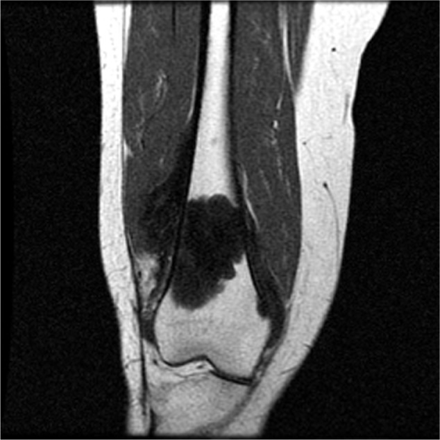
Note: Coronal T1-weighted magnetic resonance image shows marrow replacement of distal femur and a soft tissue mass extending beyond the bone cortex.
Abbreviation: MRI, magnetic resonance imaging.

Distal femur MRI.
Note: Coronal T1-weighted magnetic resonance image shows marrow replacement of distal femur and a soft tissue mass extending beyond the bone cortex.
Abbreviation: MRI, magnetic resonance imaging.