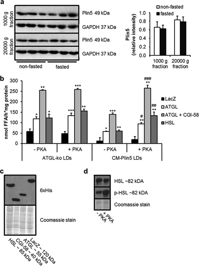
FIGURE 2.
PKA stimulates FA release from cardiac LDs of CM-Plin5 mice. a, immunoblot and densitometric analysis of Plin5 protein levels in heart homogenates (1000 × g and 20,000 × g supernatant fraction) derived from 14-week-old nonfasted and fasted WT mice. GAPDH served as loading control and cytosolic marker protein. Immunoblots are representative for two individual tissue preparations using 20 μg of protein (n = 3). b, FA release from LDs prepared from CM of ATGL-deficient and CM-Plin5 mice incubated with COS-7 cell lysates containing LacZ, ATGL, ATGL + CGI-58, and HSL in the absence or presence of PKA (−PKA/+PKA) (n = 3). Data are shown as mean ± S.D. *, p < 0.05; **, p < 0.01, and ***, p < 0.001 versus LDs incubated with LacZ containing lysates (control); #, p < 0.05; ##, p < 0.01; and ###, p < 0.001 versus CM-Plin5 LDs without PKA. c, immunoblot analysis showing protein levels of recombinant HSL, CGI-58, ATGL, and LacZ in cell lysates of transfected COS-7 cells. Coomassie stain served as loading control. d, immunoblot analysis of phosphorylated versus nonphosphorylated HSL protein levels in COS-7 cell lysates in the absence and presence of PKA. 10 μg of protein were resolved by SDS-PAGE (c and d).

