FIGURE 3.
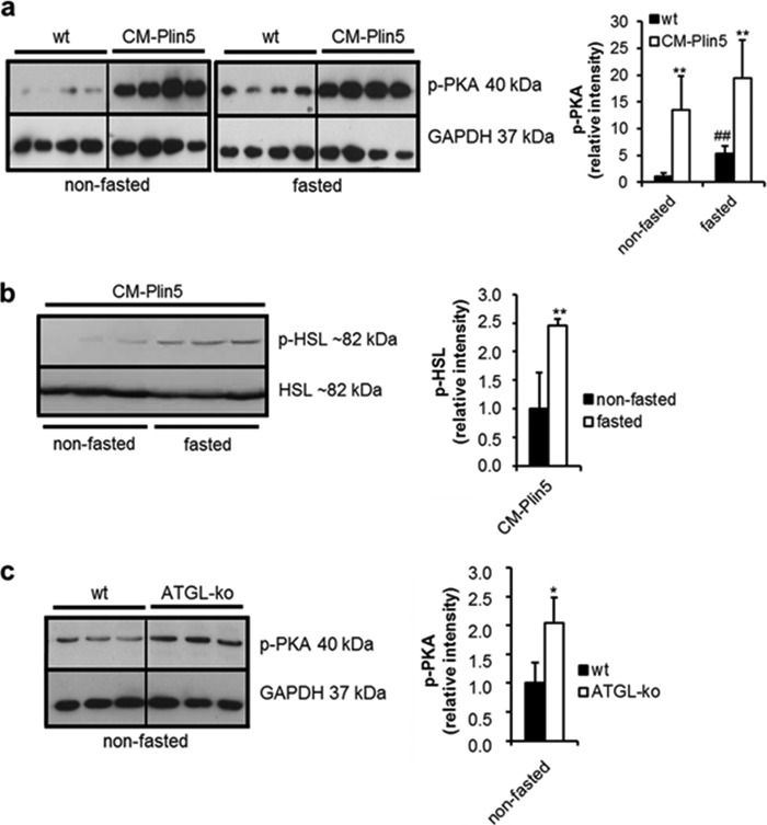
Phosphorylated PKA protein levels are highly increased in heart homogenates of CM-Plin5 mice. a, immunoblot and densitometric analysis of phosphorylated PKA (p-PKA) protein levels in heart homogenates of 12-week-old nonfasted compared with fasted WT and CM-Plin5 mice, respectively. Data are shown as mean ± S.D. *, p < 0.05, and **, p < 0.01 compared with WT; ##, p < 0.01 compared with nonfasted state (n = 4). b, immunoblot and densitometric analysis of phosphorylated HSL (p-HSL) versus nonphosphorylated HSL protein levels in cardiac lysates derived from nonfasted and fasted CM-Plin5 mice. Data are shown as mean ± S.D. **, p < 0.01 versus nonfasted state (n = 3). c, Western blot and densitometric analysis of p-PKA protein levels in cardiac homogenates of 8–11-week-old WT and ATGL-KO mice in the nonfasted state (n = 3). a–c, 20 μg of tissue protein were loaded onto the gels. Immunoblots are representative for two individual tissue preparations.

