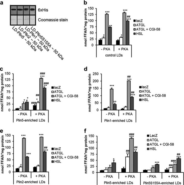
FIGURE 4.
Plin5 barrier function is under regulation of PKA. LDs were prepared from COS-7 cells transfected with Plin1-, Plin2-, Plin5-, and Plin5S155A and as control LacZ. Transfected cells were loaded with oleic acid and 3H-labeled oleic acid as tracer prior to LD isolation. a, immunoblot analysis of the LD fractions showing expression of the respective perilipin protein family member (10 μg of protein was loaded per lane). b–f, LD preparations were incubated with LacZ, ATGL, ATGL, and CGI-58 or HSL-containing COS-7 cell lysates and the release of 3H-labeled FAs was measured from control LDs (b), LDs enriched with Plin5 (c), Plin1 (d), Plin2 (e), or Plin5S155A (f). Data are mean ± S.D. of n = 3. *, p < 0.05; **, p < 0.01, and ***, p < 0.001 versus LacZ control, and ##, p < 0.01, and ###, p < 0.001 versus substrate without PKA.

