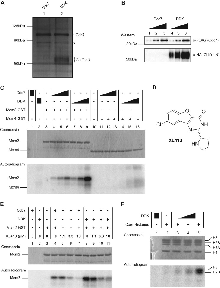
FIGURE 5.
Drosophila Cdc7 phosphorylates Mcm2 and histone H3, Chiffon stimulates Cdc7 kinase activity, and XL413 inhibits Cdc7 kinase activity. A, silver-stained SDS-polyacrylamide gel showing purified recombinant Drosophila Cdc7 or DDK complex. Cdc7 and ChiffonN were expressed in Sf21 cells as fusion proteins with N-terminal epitope tags as follows: His-FLAG, Cdc7; HA, ChiffonN. Cdc7 was expressed alone or co-expressed with ChiffonN. Cdc7 was purified by sequential Ni-NTA-agarose chromatography followed by FLAG-agarose chromatography, and DDK was purified using sequential nickel-agarose chromatography, followed by HA-agarose chromatography. Lane 1, Cdc7 purification; lane 2, Cdc7 + ChiffonN (DDK) co-purification. ChiffonN appears as a diffuse band (brace) due to phosphorylation by Cdc7. A nonspecific contaminant present at low levels in Cdc7 and DDK purifications is marked by an asterisk. B, Western analysis of Cdc7 and DDK used for kinase assay in C. Increasing amounts of Cdc7 (6, 12, or 24 ng) or DDK (6, 12, 24 ng of Cdc7, with co-purified ChiffonN) were resolved by SDS-PAGE and detected by Western blotting with antibodies against FLAG (Cdc7) and HA (ChiffonN). C, Chiffon stimulates Cdc7 kinase activity. In vitro kinase assays were performed in the presence of [γ-32P]ATP at 30 °C for 30 min. Increasing amounts of Cdc7 (6, 12, and 24 ng) or DDK complex (6, 12, and 24 ng of Cdc7, with co-purified ChiffonN) were incubated with 0.5 μg of Mcm2(N1–279)-GST or Mcm4(N1–233)-GST. Reactions were separated by SDS-PAGE, stained with Coomassie (upper panel), and exposed to a phosphor screen (lower panel). D, chemical structure of the Cdc7-specific inhibitor XL413. E, Cdc7 kinase activity is inhibited by the Cdc7-specific inhibitor XL413. Kinase assays were performed as in C. Cdc7 (12 ng) or DDK (12 ng Cdc7) was incubated with [γ-32P]ATP and 0.5 μg of Mcm2-GST in the presence of either vehicle (DMSO) or increasing concentrations of XL413 (1.1, 3.3, and 10 μm, respectively). F, DDK phosphorylates histones H3 and H2B. Increasing amounts of DDK (6, 12, and 24 ng of Cdc7) were incubated with 2 μg of core histones in the presence of [γ-32P]ATP at 30 °C for 60 min. Reactions were separated and analyzed as in C.

