
FIGURE 1.
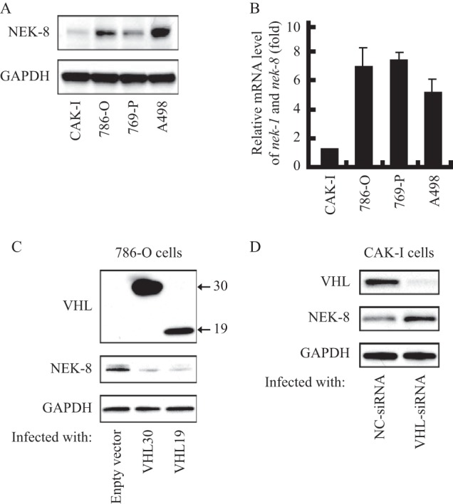
pVHL down-regulated NEK8. A, NEK8 protein expression profiles in renal cancer cells. GAPDH was employed as a loading control. B, nek8 mRNA levels in renal cancer cells. Basal levels of nek8 were measured by real-time PCR (with normalization relative to GAPDH levels). Data shown are fold-change relative to the nek8 levels in CAK-I cells. C, overexpression of pVHL leads to NEK8 degradation. 786-O cells were transfected with pcDNA3.0-VHL30, pcDNA3.0-VHL19, or empty vector to collect whole cell lysate 48 h later for immunoblot analysis of pVHL and NEK8, GAPDH was employed as a loading control. D, pVHL knockdown un-regulated NEK8. CAK-I cells were transfected with VHL-siRNA or negative control siRNA to collect whole cell lysate 48 h later for immunoblot analysis of pVHL and NEK8, GAPDH was employed as a loading control.