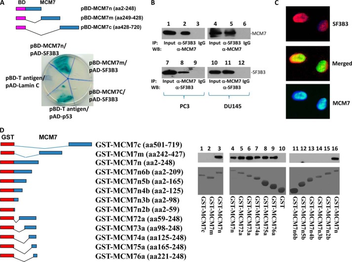
FIGURE 1.
MCM7 interacts with SF3B3. A, yeast two-hybrid analysis of MCM7 N terminus binding with SF3B3. Top panel, schematic diagram of DNA-binding domain (BD)-MCM7 fragment constructs. Bottom panel, binding of BD-MCM7 fragments with activation domain (AD)-SF3B3. Co-transfection of pBD-T-antigen and pAD-Lamin C is negative control, whereas co-transfection of pBD-T-antigen and pAD-p53 is a positive control. B, co-immunoprecipitation of MCM7 and SF3B3 proteins. Protein extracts from PC3 and DU145 cells were immunoprecipitated by the indicated antibodies and immunoblotted with antibodies specific for MCM7 (top panel) or SF3B3 (bottom panel). C, SF3B3 co-localized with MCM7. PC3 cells were stained with antibodies against MCM7 (mouse monoclonal and FITC-conjugated donkey anti-mouse antibodies) and SF3B3 (rabbit polyclonal and Cy5-conjugated donkey anti-rabbit antibodies). D, MCM7 binds with SF3B3 in cell free system. Left panel, schematic diagram of GST-MCM7 constructs. Right panel, binding of GST-MCM7 fragments with His tag-SF3B3 generated from E. coli. IP, immunoprecipitation; WB, Western blot.

