
FIGURE 5.
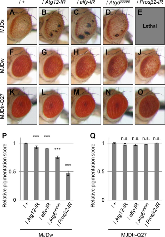
Loss of autophagic or proteasomal function causes exacerbation of eye degeneration in MJDtr-Q78 flies. A–O, light microscopic images of the external compound eyes of 1-day-old adult flies of two different MJDtr-Q78 fly lines, MJDtr-Q78S flies (A, MJDs) and MJDtr-Q78W flies (F, MJDw); MJDtr-Q78 flies co-expressing Atg12-IR (B and G), alfy-IR (C and H), or the proteasome β2 subunit (Prosβ2)-IR (E and J) or bearing an Atg6 mutation (Atg600096) (D and I); and the control 1-day-old adult MJDtr-Q27 flies (K–O). Note that Prosβ2 knockdown showed a lethal phenotype in the MJDtr-Q78S flies. Fly genotypes were as follows: gmr-GAL4/+;UAS-MJDtr-Q78S/+ (A); gmr-GAL4/+;UAS-MJDtr-Q78S/UAS-Atg12-IR (B); gmr-GAL4/+;UAS-MJDtr-Q78S/alfy-IR (C); gmr-GAL4/+;UAS-MJDtr-Q78S/+;Atg600096/+ (D); gmr-GAL4/+;UAS-MJDtr-Q78S/+;Prosβ2-IR/+ (E); gmr-GAL4/+;;UAS-MJDtr-Q78W/+ (F); gmr-GAL4/+;UAS-Atg12-IR/+;UAS-MJDtr-Q78W/+ (G); gmr-GAL4/+;UAS-alfy-IR/+;UAS-MJDtr-Q78W/+ (H); gmr-GAL4/+;;UAS-MJDtr-Q78W/Atg600096 (I); gmr-GAL4/+;;UAS-MJDtr-Q78W/Prosβ2-IR (J); gmr-GAL4/+;;UAS-MJDtr-Q27/+ (K); gmr-GAL4/+;UAS-Atg12-IR/+;UAS-MJDtr-Q27/+ (L); gmr-GAL4/+;alfy-IR/+;UAS-MJDtr-Q27/+ (M); gmr-GAL4/+;;UAS-MJDtr-Q27/Atg600096 (N); and gmr-GAL4/+;;UAS-MJDtr-Q27/Prosβ2-IR (O). P and Q, quantitative imaging analyses of eye pigmentation in the 1-day-old adult MJDtr-Q78W flies (P) or the control 1-day-old adult MJDtr-Q27 flies (Q) expressing Atg12-IR, alfy-IR, or Prosβ2-IR or bearing an Atg6 mutation. More than four eye images were analyzed for each genotype. Data are presented as the mean ± S.E. (error bars) (***, p < 0.001; n.s., not significant, versus the MJDtr-Q78W flies in P and versus the MJDtr-Q27 flies in Q, respectively).