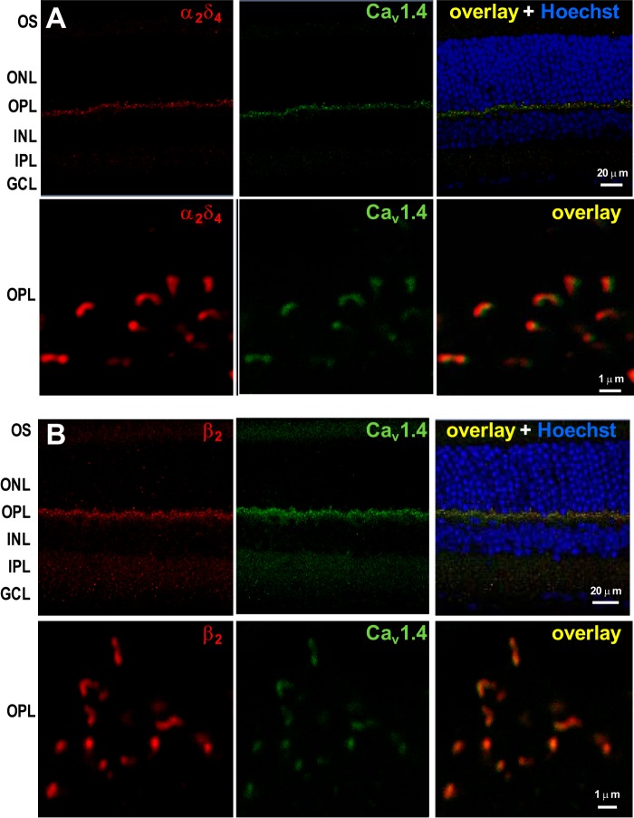
FIGURE 4.
Colocalization of Cav1.4 α1 with β2 or α2δ4 at the photoreceptor synaptic ribbon. Mouse retina sections were double-labeled with anti-Cav1.4 α1 (green) and anti-α2δ4 (red) (A) or anti-β2 (red) (B). The overlay is shown in the right panel together with Hoechst staining. Higher magnification images of the OPL are shown in the bottom panels. Cav1.4 shows a high degree of colocalization with β2 and α2δ4, as indicated by the calculated Pearson's correlation coefficient between the Alexa 488-stained Cav1.4 and the Alexa 555-stained β2 (r ≥ 0.710) or α2δ4 (r ≥ 0.608). OS, outer segment; INL, inner nuclear layer; IPL, inner plexiform layer; GCL, ganglion cell layer.

