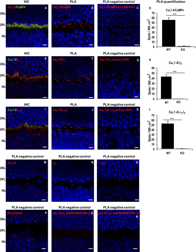
FIGURE 6.
Cav1.4 α1 interacts with CaBP4, β2 and α2δ4 in mouse retina. Immunohistochemistry (IHC) or PLAs were performed in sections of WT or knock-out mouse retina using anti-Cav1.4 α1 and anti-CaBP4 (A–D), anti-Cav1.4 α1 and anti-β2 (E–H), anti-Cav1.4 α1 and anti-α2δ4 (I–L), anti-Cav1.4 (M), anti-β2 (N), anti-α2δ4 (O), no primary antibodies (P), anti-β2 preadsorbed with β2a (Q), or anti-α2δ4 preadsorbed with α2δ4 (R). For IHC, WT mouse retina sections were double-labeled with anti-Cav1.4 α1 (red) and in green anti-CaBP4 (A), anti-Cav1.4 α1 (green) and in red anti-β2 (E), or anti-α2δ4 (I). INL, inner nuclear layer. PLA was performed on retina from WT (B, F, J, and M–R), CaBP4 KO (C), or Cav1.4 KO (G and K) mice. Sites of protein interaction are visualized as red spots. Quantification of PLA spots are shown as mean ± S.E. (error bars) for Cav1.4/CaBP4 in WT (n = 7) versus CaBP4 KO mice (n = 3) (p < 0.001; Student's t test) (D), for Cav1.4-β2 in WT (n = 7) versus Cav1.4 KO mice (n = 3) (p < 0.001) (H), and for Cav1.4-α2δ4 in WT (n = 6) versus Cav1.4 KO mice (n = 3) (p < 0.001) (L). For all images, nuclei are labeled with Hoechst. Scale bar, 5 μm.

