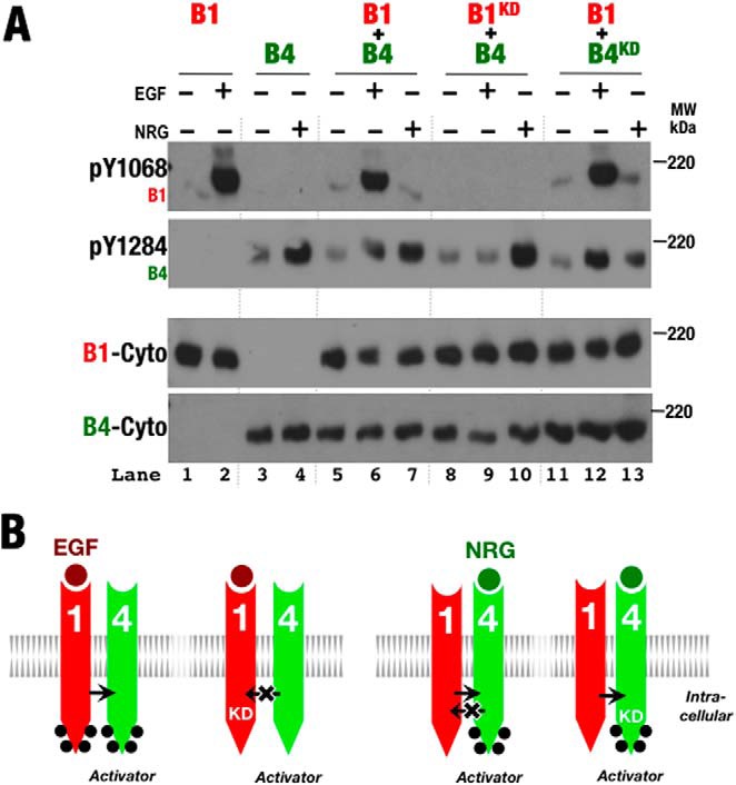
FIGURE 7.

ErbB4 preferentially serves as the activator kinase when paired with ErbB1. A, Western blots of whole cell lysates from S2R+ cells transiently transfected with wild-type and kinase-deficient (KD) forms of ErbB1 (B1) and ErbB4 (B4) and treated with EGF or NRG1β (+) or left untreated (−). Antibodies against phospho-Tyr1068 (pY1068; ErbB1), phospho-Tyr1284 (pY1284; ErbB4), and the ErbB1 (B1-Cyto) and ErbB4 (B4-Cyto) cytoplasmic regions were used as indicated. B, schematic representation of phosphorylation patterns observed in A. Phosphorylation is indicated by black dots.
