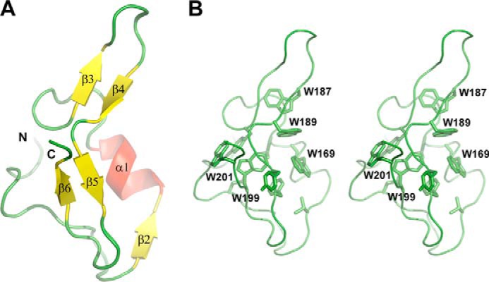
FIGURE 3.

Structure of the MG200 EAGR box domain (Protein Data Bank code 4DCZ). A, schematic representation of the crystal structure of the MG200 EAGR box. The secondary structure element nomenclature is consistent with that previously reported (28). B, stereo view of the MG200 EAGR box with all of the tryptophan residues from the hydrophobic core depicted in stick representations.
