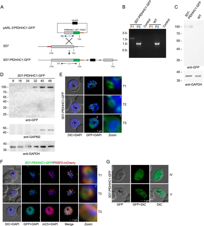
FIGURE 2.
PfDHHC1 is an IMC localized palmitoyl acyltransferase. A, schematic representation of the GFP replacement of the endogenous 3′ end of PfDHHC1 creating a 3D7-PfDHHC1-GFP cell line. The vector encompasses the selection cassette (black), and 1 kb of the PfDHHC1 gene (gray) was fused to GFP (green) accompanied by the 3′UTR of PbDT (orange) without any promoter. Integration of the vector takes place by homologous recombination (cross) into the PfDHHC1 locus creating a full-length PfDHHC1-GFP fusion under the control of the endogenous promoter. B, integration was confirmed by PCR using two different primer combinations on gDNA. One primer set (red) hybridizes in a PfDHHC1 region upstream of the integration and in the coding region of GFP. This primer combination can only amplify a 1.7-kb DNA fragment after recombination took place. The other set (blue) amplifies 1.1 kb of PfDHHC1 in the parental as well as in the transgenic parasite line. Control indicates PCRs with the red primer set in the absence of parasite DNA. C, expression of the transgene from the endogenous locus was shown by Western blot analysis using anti-GFP antibodies (upper panel) resulting in a fusion protein of ∼100 kDa (calculated mass of 100 kDa) using late stage parasite material. Anti-GAPDH was used as a loading control. D, stage-specific expression pattern of PfDHHC1-GFP using synchronized 3D7-PfDHHC1-GFP parasite material (harvested after 8, 16, 24, 32, 40, and 48 h) and anti-GFP antibodies. Antibodies directed against the IMC marker protein GAPM2 were used as a stage-specific control. Anti-GAPDH antibodies were used as loading control. E, PfDHHC1-GFP was localized in unfixed late stage parasites showing characteristic IMC dynamics during schizogony (T1 to T3). Nuclei stained with DAPI (blue). Enlargement of selected areas are marked with a white square and referred to as zoom. Scale bar, 1 μm. F, co-localization of PfDHHC1-GFP and PfISP3mCherry. PfISP3mCherry was episomally co-expressed and shows identical spatial distribution compared with PfDHHC1-GFP. G, PfDHHC1-GFP distribution within the nascent IMC during gametocytogenesis (stage IV and V). These symmetric sutures of the IMC are expanding with ongoing growth of the IMC vesicles and maturation of the gametocytes (stage IV and V). DIC, differential interference contrast. Scale bars, 1 μm.

