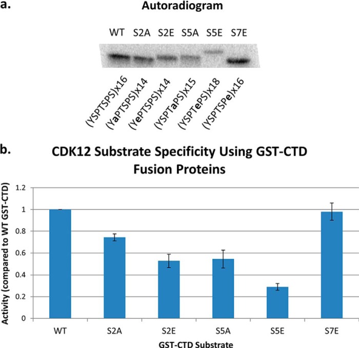
FIGURE 6.
Phosphorylation of mutant GST-CTD substrates by hCDK12/CyclinK. a, representative autoradiogram of a SDS-PAGE gel from an hCDK12/CyclinK kinase assay using mutant GST-CTD substrates. The composition and number of the CTD heptad repeats in the GST-CTD fusion protein is indicated below the appropriate lanes. b, quantification of the degree of phosphorylation of each mutant substrate using a PhosphorImager and ImageQuant software (normalized to a value of 1.0 for the wild-type GST-CTD fusion protein). Error bars are ± 2 S.E., n = 3.

