Fig.3.

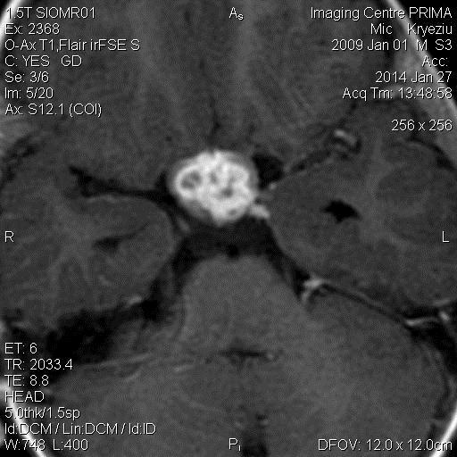
Post-gadolinium enhanced T1W images in axial section showing multiple ring enhancing lesions clustered around optic chiasma in the suprasellar cistern

Post-gadolinium enhanced T1W images in axial section showing multiple ring enhancing lesions clustered around optic chiasma in the suprasellar cistern