. Author manuscript; available in PMC: 2015 Feb 25.
Published in final edited form as: Spine (Phila Pa 1976). 2014 Feb 1;39(3):E166–E173. doi: 10.1097/BRS.0000000000000073

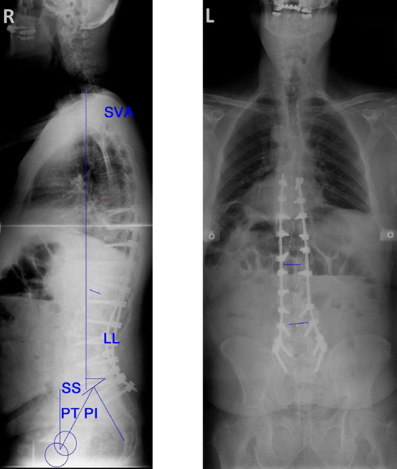
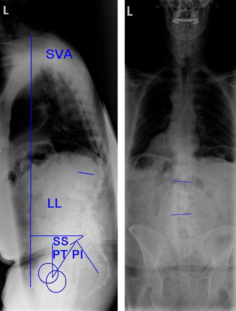
Figure 1.

Figure 1

Figure 1

Preoperative and postoperative full–length coronal and sagittal standing radiographs for a 66–year old male patient diagnosed with progressive degenerative scoliosis. He had a surgical history of posterior in situ fusion (L4–Sacrum). He underwent circumferential spine surgery with a total of eight lateral interbody fusion levels and percutaneous posterior fusion using pedicle screws (T8–Sacrum).

Coronal Cobb angle (°) SVA LL PT SS PI

Preoperative 15° 13 cm 43° 33° 34° 67°

Postoperative 4.1 cm 57° 29° 35° 64°

Sagittal Vertical Alignment (SVA), Lumbar Lordosis (LL), Pelvic Tilt (PT), Sacral Slope (SS), and Pelvic Incidence (PI).