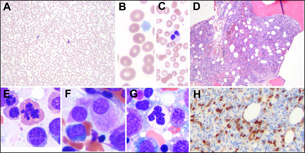
Fig 1.
Morphological features of a typical case of MDS. (A–C) The peripheral smear shows severe pancytopenia (A), with a macrocytic anemia (B), neutropenia with dysplastic neutrophils and thrombocytopenia with pale platelets (C). (D) The marrow is typically hypercellular, indicating ineffective hematopoiesis. (E–H) The marrow cytology reveals dysplasia in the erythroid (E), megakaryocytic (F), and granulocytic lineage (G), as well as increased blasts, which can sometimes be detected by CD34 immunostaining (H).

