Figure 7.
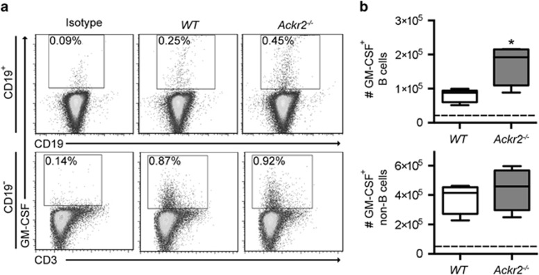
Ackr2 deficiency leads to increased numbers of GM-CSF+ B cells in draining LNs after immunization with MOG1–125 protein. EAE was induced in WT littermate control (WT) and Ackr2-deficient (Ackr2−/−) C57BL/6 mice (n=4) by immunization with MOG1–125 protein (50 μg in 3 mg ml−1 Mycobacterium tuberculosis H37RA CFA), draining LNs harvested on day 8, and the number of GM-CSF+ CD19+ B cells determined. (a) Representative flow cytometry plots that were pregated on live IgD+IgM+ singlets (top panels) and live IgD−IgM−CD19− singlets (bottom panels), and the percentage of GM-CSF+ CD19+ B cells and GM-CSF+ CD19− non-B cells respectively are shown. (b) Box and whisker plots, where the boxes represent the 25th to 75th percentiles, the lines within the boxes represent the median and the lines outside the boxes represent the 5th and 95th percentiles, show the absolute number of GM-CSF+ B cells (top plot) and GM-CSF+ non-B cells (bottom plot) in the draining LNs. The dotted line represents the isotype control level. A repeat experiment yielded similar results. *P<0.05 by Mann–Whitney test.

