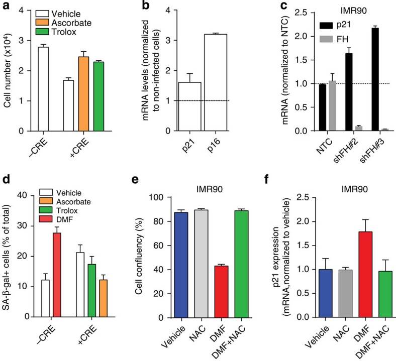
Figure 6. GSH succination leads to redox stress-induced senescence in primary kidney cells and human diploid fibroblasts.
(a) Primary epithelial kidney cells (1 × 10⁴) from Fh1fl/fl animals were either left uninfected or infected with adeno-CRE and treated with vehicle or the antioxidant trolox or ascorbate. Cell number was measured after 2 days of culture. (b) The ablation of FH in cells described in a resulted in the activation of p16 and p21 transcription. Results are expressed as average fold induction over non-infected cells±s.e.m (representative experiments; n=6 for p21 and n=3 for p16). (c) FH and p21 mRNA levels were assessed by qPCR in non-targeting control (NTC)-transfected cells in FH-silenced cells. (d) Microscopy quantitative analysis of senescence-associated β-galactosidase activity, induced by DMF or adeno-CRE infection, with or without trolox and ascorbate. (e,f) cell growth (e) and p21 mRNA expression levels (f) were assessed in IMR90 cells after DMF treatment, with or without the antioxidant N-acetyl cysteine (NAC). All the results were obtained from three independent cultures and expressed as average±s.e.m.

