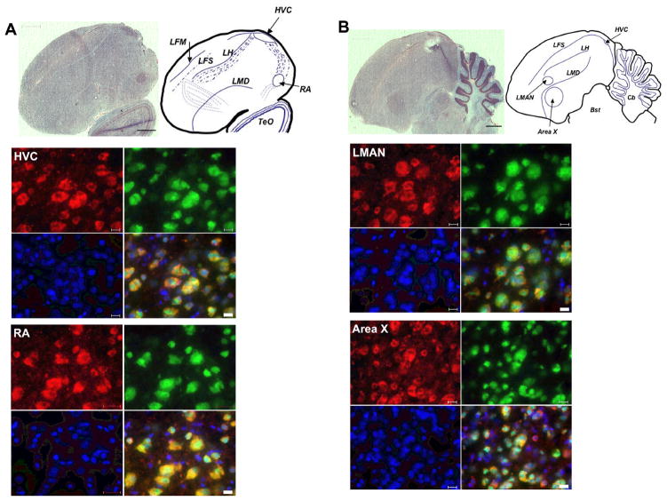
Fig. 2. FMRP is expressed in neurons in the male zebra finch brain.
Sagital brain sections containing A. HVC (letter-based name) and RA and B. LMAN (Lateral Magnocellular nucleus of the Anterior Nidopallium) and Area X were stained for anatomy with cresyl violet (bar = 1000μm) (upper left). Upper right is an accompanying sketch with the significant anatomical features indicated. Adjacent sections were co-stained with the zebra finch-specific Fmrp antibody 24 (red), the neuronal marker NeuN (green), and DAPI (blue). In the overlay, a yellow signal indicates co-fluorescence for red and green. Bar = 20μm. Bst: Brainstem; Cb: Cerebellum; LFM: Lamina frontalis suprema; LFS: Lamina frontalis superior; LH: Lamina hyperstriatica; LMD: Lamina medullaris dorsalis; TeO: Optic Tectum. Shown are images from a posthatch day (P) P30 brain; P60 and Adult males showed similar results (data not shown). [Reprinted with permission from (Winograd et al., 2008).]

