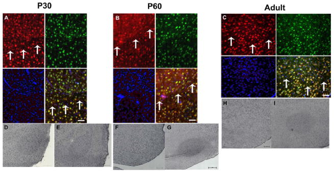
Fig. 3. FMRP is consistently elevated in the RA nucleus of a P30 male zebra finch and variably expressed in P60 and Adult males.

A–C. Representative fluorescent-immunohistochemistry using an antibody specific to zebra finch FMRP on a male P30 (A) P60 (B) and Adult (C) zebra finch RA. Shown are FMRP immunoreactivity (red), NeuN stain (green), and DAPI-labeled nuclei (blue), along with the overlay. Arrows denote ventral border of RA. Bar = 100μm. D–I. Representative DAB-IHC using anti-zebra finch FMRP antibody on a male P30 (D, E) P60 (F, G) and Adult (H, I) zebra finch RA. Bar = 200μm. [Reprinted with permission from (Winograd et al., 2008).]
