
Figure 7.
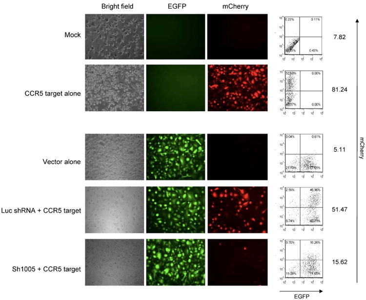
Specific knockdown of mCherry-CCR5 target expression by sh1005 in macrophages derived from FL-CD34+ cells. FL-CD34+ cells (2 × 10⁵) were transduced with lentiviral vectors expressing sh1005 or Luc shRNA. The cells were harvested 3 days after virus transduction, sorted by the EGFP expression, and further transduced with the lentiviral vector encoding mCherry-CCR5 target sequence at an MOI of 0.5. FL-CD34+ cells were then differentiated to macrophages with 20 ng/ml of IL-3, 50 ng/ml of SCF and 20 ng/ml of MCSF for 9 days and then with 5 ng/ml of GM-CSF for 5 days. The effect of shRNA was monitored by the expressions of mCherry by florescence microscopy and by flow cytometry. The results are exhibited as mCherry dot plots versus EGFP dot plots. The number in each quadrant represents the percentage in each population. The numbers on right side of each panel show the mean fluorescent intensity of the mCherry expression