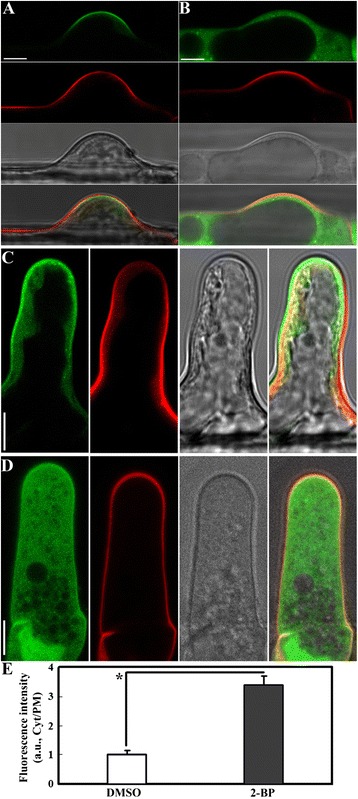
Figure 5.

2-BP treatment re-distributed the PIP 2 sensor from the PM to the cytoplasm in root hairs. A. DMSO-treated root hairs expressing the PIP2 sensor (green) at the initiating stage. B. 2-BP-treated root hairs expressing the PIP2 sensor at the initiating stage. C. DMSO-treated root hairs expressing the PIP2 sensor at the elongating stage. D. 2-BP-treated root hairs expressing the PIP2 sensor at the elongating stage. E. Ratio of fluorescence signals. a.u. stands for arbitrary fluorescence units. Cyt/PM indicates the ratio of cytoplasmic to the plasma membrane signal. Results are means ± standard deviation (SD, n = 30). Asterisk indicates significant difference (Student’s t-test, P < 0.01). Root hairs were stained with the fluorescence dye propidium iodide (red) to outline cell shape. Corresponding bright-field images are shown together with merges of different channels. Bars = 7.5 μm.
