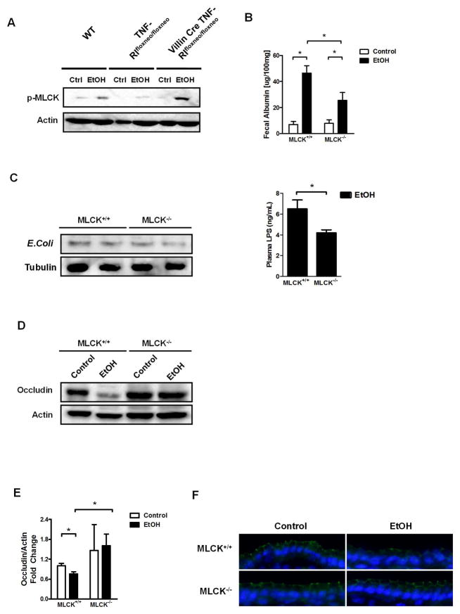
Figure 6. MLCK is involved in TNFα signaling and contributes to alcohol-induced barrier loss.
(A) Wild type (WT), TNFRIflxneo/flxneo and VillinCre TNFRIflxneo/flxneo male mice were treated with ethanol or dextrose as control (ctlr) by gavage once (n = 3–4). Representative western blot for phosphorylated MLCK (p-MLCK) in epithelial cells isolated from the jejunum. (B–F) MLCK+/+ and MLCK−/− littermate mice were orally fed a control (n = 4) and alcohol diet (n = 10–14). (B) Fecal albumin content. (C) Western blot for E. Coli proteins in liver (left panel); plasma LPS level (right panel). (D) Representative western blot for occludin in the jejunum. (E) Occludin quantification of western blots. (F) Representative immunofluorescent staining for occludin; nuclei are stained blue. *p < 0.05.

