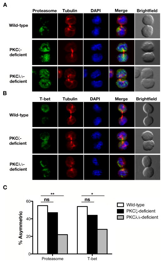
Figure 2.
Loss of PKCζ or PKCλ/ι impairs asymmetric segregation of proteasome and T-bet during the first division of an activated naïve CD8+ T cell. Confocal microscopy of (A) proteasome or (B) T-bet (green), β-tubulin (red), and DNA (blue; stained with the DNA-intercalating dye DAPI) in sorted wild-type, PKCζ-deficient, or PKCλ/ι-deficient OT-I CD8+ T cells undergoing their first division after adoptive transfer into Lm-OVA infected recipient mice. (C) Incidence of asymmetric protein localization from T cells shown in (A) and (B). The number of dividing cells from two experiments is indicated in parenthesis as follows (wild-type, PKCζ-deficient, PKCλ/ι-deficient): proteasome (58, 30, 27) and T-bet (28, 34, 43). ns, not significant; *P < 0.05 and **P < 0.01 (one-tailed unconditional Fisher’s exact test).

