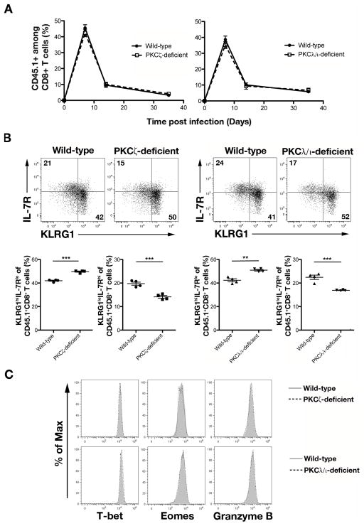
Figure 4.
PKCζ- and PKCλ/ι-deficient CD8+ T cells give rise to reduced KLRG1lo IL-7Rhi T cells at day 7 post-infection. (A) Percentages of CD45.1+ cells of CD8+ T cells on days 7, 14, and 35 in the blood of mice that received 5×10³ wild-type (closed circles) or PKCζ-deficient (open squares) OT-I CD45.1+CD8+ T cells (left) or wild-type (closed circles) or PKCλ/ι-deficient (open squares) OT-I CD45.1+CD8+ T cells (right) and were infected with Lm-OVA; points represent mean ± SEM; n ≥ 4/group. (B) Expression of KLRG1 and IL-7R by CD45.1+CD8+ T cells in the spleen on day 7 post-infection (top). Frequencies of KLRG1hiIL-7Rlo and KLRG1loIL-7Rhi cells (bottom); each point represents an individual mouse and lines indicate the mean ± SEM; n = 4/group. **P < 0.01 and ***P < 0.001 (two-tailed unpaired t-test). (C) Expression of T-bet, Eomes, and Granzyme B by wild-type (gray filled) and PKCζ-deficient (dashed black) CD45.1+CD8+ T cells (top) or wild-type (gray filled) and PKCλ/ι-deficient (dashed black) CD45.1+CD8+ T cells (bottom) in the spleen on day 7 post-infection. Data are representative of three experiments.

