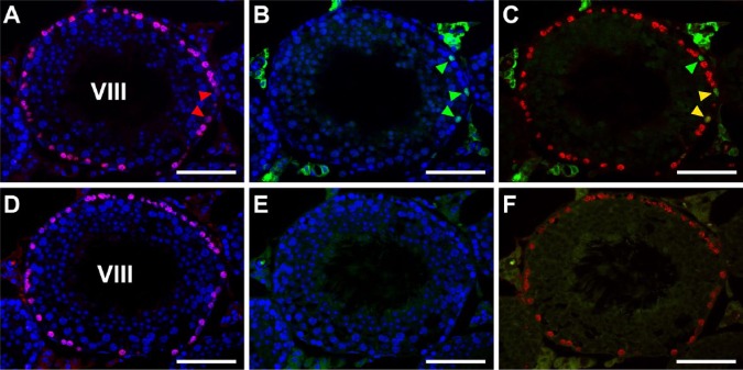
Figure 4.
Fluorescence images of mouse seminiferous tubules showing the effects of different pH conditions of heat-induced antigen retrieval (HIAR) on double immunostaining of BrdU (red) and PLZF (green). Nuclei were counterstained with Hoechst 33258 (blue). Paraffin sections of the testis were pretreated with HIAR with 20 mM Tris-HCl buffer, pH 9.0 (A–C) or 10 mM citrate buffer, pH 6.0 (D–F), and were then immunostained with mouse monoclonal anti-BrdU antibody together with the rabbit polyclonal anti-PLZF antibody. (A–C) and (D–F) represent two serial sections of the same stage VIII seminiferous tubule. Pictures in different combinations of wavelengths are shown. The immunoreactivity of BrdU (red) was localized in the nuclei of numerous cells (including two cells indicated by red arrowheads) that were in contact with the basement membrane, with similar intensities observed between the pretreatment in pH 9.0 (A-–C) and that in pH 6.0 (D–F). With pH 9.0, the immunoreactivity of PLZF (green) was clearly detected in the nuclei of three cells (green arrowheads, B). Two of the PLZF-positive cells were simultaneously positive for BrdU (yellow arrowheads), whereas the other was negative for BrdU (green arrowhead) in the merged picture (C). However, at pH 6.0, the immunoreactivity of PLZF was hardly detected in any cells, and there was high background fluorescence in the cytoplasm of elongated spermatids (E, F). Scale, 50 μm.

