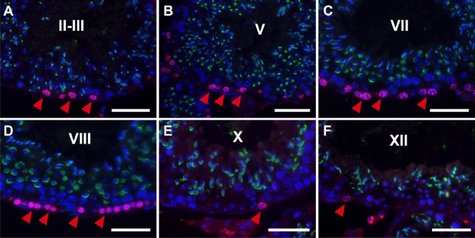
Figure 5.
Fluorescence images of mouse seminiferous tubules showing the simultaneous IHC of BrdU (red) and peanut agglutinin-lectin (PNA-LHC; green). Nuclei were counterstained with Hoechst 33258 (blue). Paraffin sections of the mouse testis were pretreated with heat-induced antigen retrieval (HIAR) and then immunostained with mouse monoclonal anti-BrdU antibody and PNA lectin. The merged images of different wavelengths are shown. At stages II-III (A), V (B), VII (C), VIII (D), X (E), and XII (F), the immunoreactivity of BrdU was localized in the nuclei of cells that are in contact with or close to the basement membrane (red arrowheads). Scale, 25 μm.

