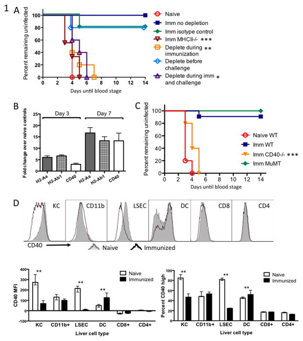
Figure 1. CD4+ T cells and CD40 are needed for protective immunity conferred by fabb/f- immunization.
(A) Groups of WT C57BL/6 mice were immunized twice with 1×10⁴ fabb/f- sporozoites then challenged one month later and monitored for blood-stage parasitemia. Each group was either depleted of CD4+ T cells just prior to each immunization, just prior to challenge, both, or left untreated. (B) Gene expression in the livers of fabb/f- immunized mice measured by q-RT-PCR. WT mice were immunized once with 5×10⁴ fabb/f- sporozoites and 7 days later RNA was isolated from a section of whole liver. Values were normalized to Gapdh and are shown as fold-change over naïve control liver. (C) Groups of WT or gene-deficient C57BL/6 mice were immunized twice with 1×10⁴ fabb/f- sporozoites then challenged one month later and monitored for blood-stage parasitemia. (D) Kupffer cells (KC), CD11b+ infiltrating myeloid cells, liver sinusoidal endothelial cells (LSEC), dendritic cells (DC), CD8+ and CD4+ T cells were isolated from the livers of fabb/f-immunized mice on Day 7 and stained for CD40 surface molecule expression. Representative samples are shown in the top panel and summarized data are shown below. The MFI, right, is based on the entire histogram and percent CD40 high, left, is based on the CD40-bright peak of the dendritic cells. Asterisks represent significant differences between naïve and immunized within one cell type.

